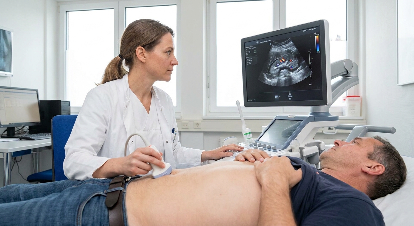
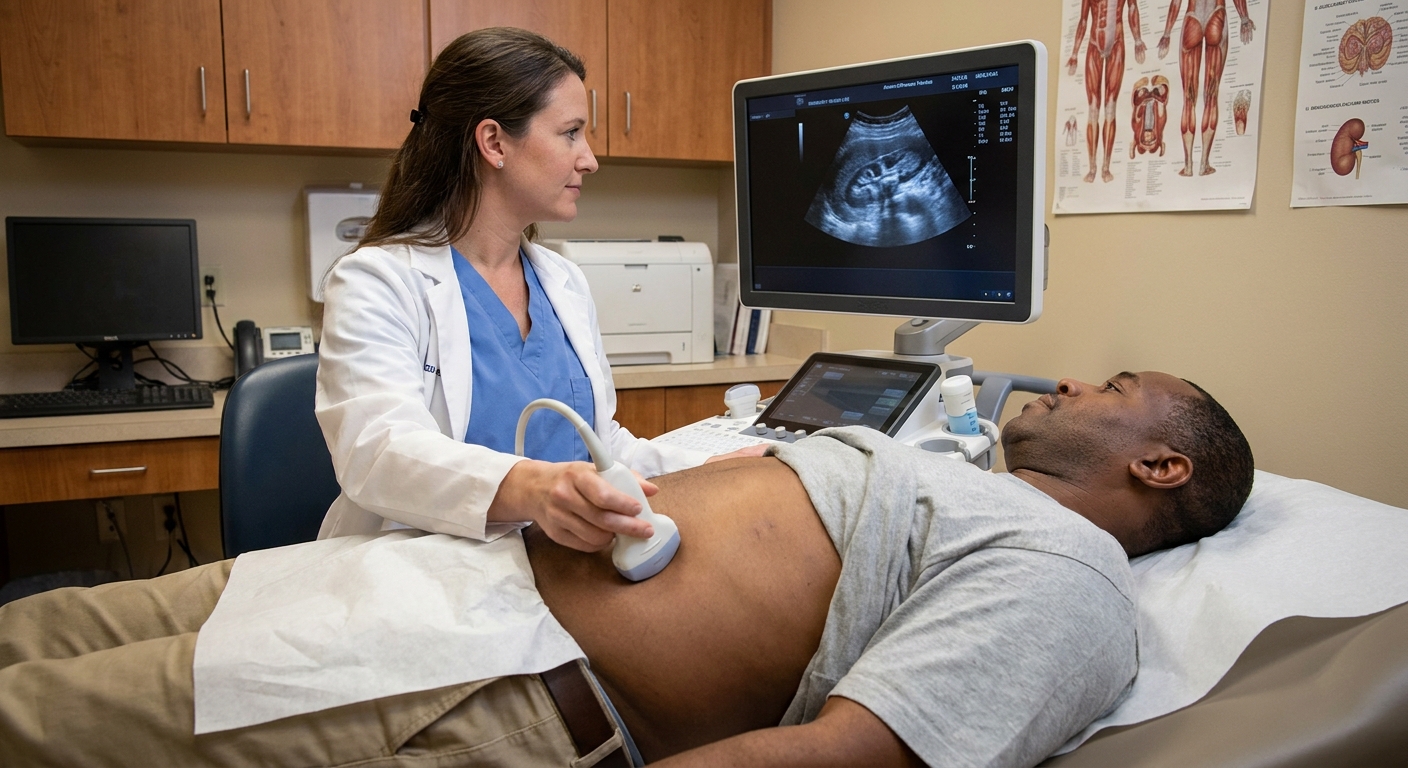
If you just read “horseshoe kidney” on an ultrasound, CT scan, or MRI report, it can sound alarming. In many cases, it is simply a different way your kidneys formed before birth. Plenty of people live their whole lives never knowing they have it. The important part is what this anatomy can change, especially how urine drains and how clinicians plan procedures.
A horseshoe kidney, explained simply
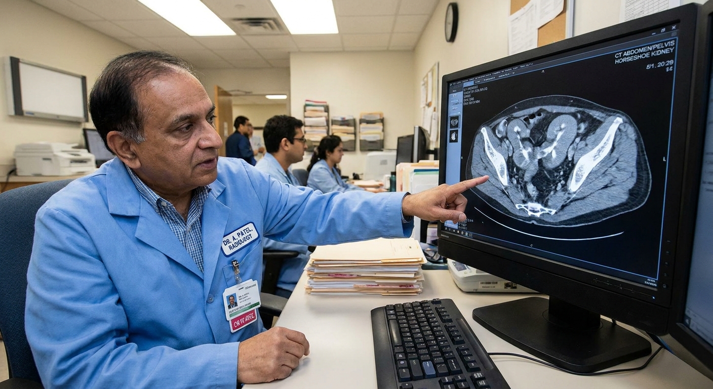
Normally, you have two separate kidneys, one on each side of your spine, sitting fairly high in the back of the abdomen. A horseshoe kidney happens when the two kidneys fuse across the midline during early development. The connection is called an isthmus, which is a bridge of kidney tissue or fibrous tissue joining the two sides.
Because of how the fused kidneys get “caught” as they move into position during fetal development, a horseshoe kidney typically sits lower than usual in the abdomen. The blood vessel anatomy can also be different, with extra or unusual arteries supplying the kidney.
Key point
A horseshoe kidney is a congenital anatomy variant. It is not something you caused, and it is not contagious. It does not automatically mean kidney failure or cancer. It does mean your kidney plumbing and positioning may be a little different, which can affect risk and medical planning.
How common is horseshoe kidney?
Horseshoe kidney is one of the more common kidney formation differences. It is often cited at roughly 1 in 400 to 1 in 600 people, and it is reported more often in males. Many people find out incidentally when they get imaging for something unrelated, like abdominal pain, a urinary tract infection workup, or a scan after an injury.
Some people have horseshoe kidney along with other urinary tract differences. If your report mentions additional findings, your clinician may recommend follow-up imaging or a referral to urology or nephrology depending on the situation.
Why urine drainage problems are more likely
The kidneys continuously make urine, which drains into a funnel-like collecting area and then into a tube called the ureter, which carries urine down to the bladder. With a horseshoe kidney, the kidney’s lower position and altered rotation can change the angle where urine drains. That can make it easier for urine to slow down or back up.
One common pinch point is where the kidney meets the ureter, called the ureteropelvic junction (UPJ). A higher chance of narrowing or kinking at that junction is one reason people with horseshoe kidney may be diagnosed with hydronephrosis (swelling of the kidney from urine buildup).
If you want more detail on UPJ obstruction, including symptoms, tests, and treatment options, see:
- What Is Ureteropelvic Junction Obstruction?
Vesicoureteral reflux (VUR)
Another condition that can occur more often with horseshoe kidney is vesicoureteral reflux (VUR), where urine flows backward from the bladder toward the ureter and kidney. VUR is diagnosed more commonly in children, but adults can also be evaluated if they have recurrent infections, scarring, or concerning imaging findings.
VUR is not something your clinician automatically assumes is present just because you have a horseshoe kidney. It is usually considered when symptoms or imaging suggest a problem, and evaluation is individualized (often through a specialist).
Kidney stones and infections
When urine does not drain smoothly, it tends to sit longer in the kidney or collecting system. Stagnant urine can contribute to:
- Kidney stones, because minerals have more time to crystallize and clump
- Urinary tract infections (UTIs), because bacteria can grow more easily when urine does not flush out well
This does not mean everyone with horseshoe kidney will get stones or recurrent infections. It does mean that if you have repeated UTIs, flank pain, blood in the urine, or known stones, your clinician may pay extra attention to whether drainage anatomy is playing a role.

Why injury risk can be different
Because a horseshoe kidney often sits lower and can be positioned a bit more toward the front than typical, it may be less protected by the rib cage. In real life, that matters most in situations like contact sports, significant falls, or car accidents.
Most people do not need special restrictions, but if you have a horseshoe kidney and you experience a significant abdominal impact followed by symptoms like flank or abdominal pain, blood in the urine, dizziness, or fainting, that is a reason to get urgent medical evaluation.
Surgery and procedure planning
One of the biggest practical implications of a horseshoe kidney is planning for procedures.
Because the kidneys are connected and may have atypical blood vessels, surgeons and interventional radiologists usually want a clear roadmap before operating in the abdomen or retroperitoneal space. This can include:
- Different locations of renal arteries and veins than expected
- The isthmus that often lies anterior to the aorta and inferior vena cava
- Ureters that pass over or near the isthmus, affecting drainage and surgical access
This does not mean surgery is impossible. It means the team may use additional imaging, choose a specific approach, or involve specialists such as urology to reduce risk.
Horseshoe kidney and duplex systems
Some people have more than one urinary tract variant at the same time. One that sometimes shows up on imaging reports is a duplex kidney, which means there can be two collecting systems or two ureters draining a kidney (partially or completely).
If your report mentions duplicated collecting system, duplex kidney, or double ureter, this page explains what that anatomy can mean and what follow-up typically looks like:
- What Is a Duplex Kidney?
Symptoms
Many people have no symptoms at all. When symptoms do happen, they are often related to urine flow issues, stones, or infections rather than the fused kidney itself.
Possible symptoms that deserve a call to your clinician
- Recurrent UTIs or burning with urination plus fever
- Flank or lower back pain, especially if severe or one-sided
- Blood in the urine (pink, red, or cola-colored urine)
- Nausea and vomiting with pain (can happen with stones or obstruction)
- New swelling of a kidney on imaging (hydronephrosis)
Cancer risk
Most people with a horseshoe kidney never develop kidney cancer. Some studies report a modestly higher relative frequency of certain tumors in people with horseshoe kidneys, including urothelial (transitional cell) cancer of the renal pelvis, and Wilms tumor in children. Even when relative risk is higher in studies, the absolute risk is still low.
The practical takeaway is simple: persistent blood in the urine, unexplained weight loss, a new mass on imaging, or ongoing symptoms that do not fit a simple UTI should be evaluated promptly, whether or not you have a horseshoe kidney.
Pregnancy
Many people with a horseshoe kidney have normal pregnancies. Because pregnancy already increases the risk of urinary stasis and UTIs, your clinician may be more alert for infection or obstruction symptoms if you have a horseshoe kidney. If you are pregnant and develop fever, flank pain, or urinary symptoms, it is worth calling your obstetric team quickly.
How it is diagnosed
Horseshoe kidney is usually diagnosed on imaging, such as:
- Ultrasound, often the first test used
- CT, which can show anatomy, stones, and obstruction clearly
- MRI, sometimes used to evaluate anatomy without radiation
- Nuclear medicine renal scan, sometimes used to evaluate how well each side functions and how urine drains
If there are concerns about obstruction, your clinician may focus less on the label “horseshoe kidney” and more on the functional question: Is urine draining normally, and are the kidneys working well?
Treatment and follow-up
There is no treatment needed for the horseshoe shape itself if kidney function is good and urine is draining well.
Care is usually focused on managing complications if they arise, such as:
- Obstruction (for example, UPJ obstruction)
- Kidney stones
- Infections
- Reflux (VUR), when present
Follow-up is individualized. Depending on your history and what was found initially, your clinician may recommend baseline or periodic checks such as kidney function labs, blood pressure, and urinalysis, plus imaging if there is hydronephrosis, stones, recurrent infections, or symptoms.
Related genetic conditions
Horseshoe kidney can be seen more often in some genetic conditions (for example, Turner syndrome and trisomy 18). For many adults who discover a horseshoe kidney incidentally, this is not a new concern and does not mean there is an underlying syndrome. If there are other findings in your personal or family history, your clinician can advise you on whether any additional evaluation is appropriate.
When to seek urgent care
Go to urgent care or the emergency department right away if you have any of the following:
- Fever with chills and urinary symptoms, or fever with flank pain
- Severe flank or abdominal pain that does not improve
- Blood in the urine with clots, or inability to urinate
- Symptoms of dehydration or shock (fainting, confusion, severe weakness)
These symptoms can happen with or without a horseshoe kidney, but the anatomy can raise the odds that an infection or blockage becomes a bigger issue.
Questions to ask at your next appointment
- Is my urine draining normally from both sides?
- Do I have hydronephrosis, and if so, how severe is it?
- Do you see stones, scarring, or signs of repeated infections?
- Do you think reflux (VUR) is a concern for me?
- Do you recommend seeing a urologist or nephrologist?
- If I ever need abdominal surgery, what should I tell the surgeon ahead of time?
FAQ
Is horseshoe kidney dangerous?
Often, no. Many people never have problems. The main concern is a higher risk of issues like obstruction, reflux, stones, or infections. The level of risk varies person to person.
Can you live a normal life with a horseshoe kidney?
Yes, many people do. If you are prone to stones or infections, you may need more monitoring and preventive habits, like staying well-hydrated and following your clinician’s guidance.
Does horseshoe kidney mean I will need surgery?
Not automatically. Surgery is only considered if there is a specific problem to fix, such as a significant obstruction or a stone that cannot pass safely.
Should my family members be tested?
Routine screening for relatives is not typically needed unless there are symptoms or other medical reasons. If there is a strong family history of kidney or urinary tract differences, your clinician can advise you on whether any evaluation makes sense.