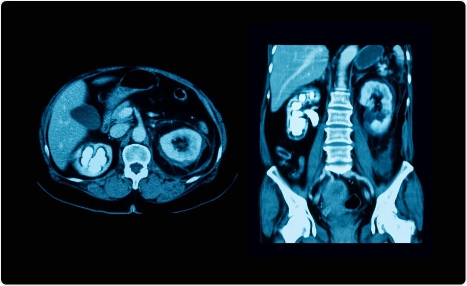
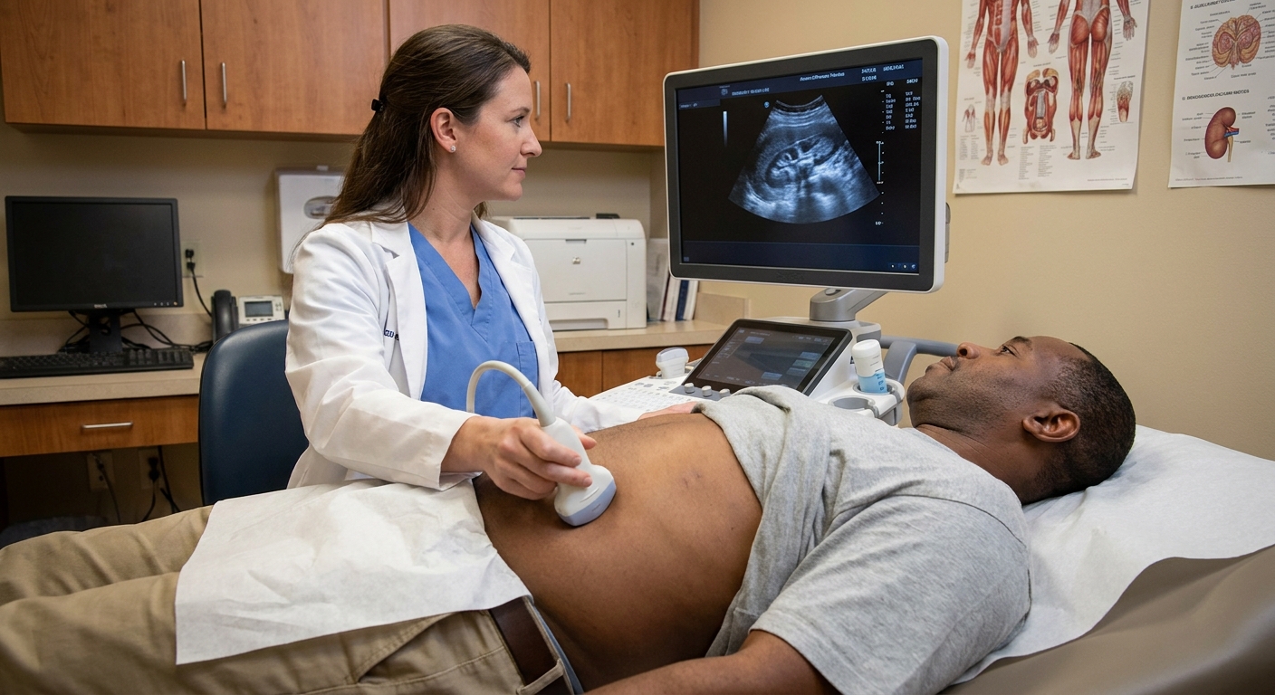
If you are reading your CT or ultrasound report and your eyes snag on the word hydronephrosis, take a breath. I have had many patients walk into clinic worried that a “swollen kidney” automatically means permanent damage. Hydronephrosis is best understood as a clue on imaging, not a final diagnosis. It means urine is not draining as smoothly as it should, so the kidney’s collecting system is stretched.
In this article, I will explain what hydronephrosis is, what the grades mean, the most common reasons urine backs up, and why your radiology report often cannot name the cause by itself.
What hydronephrosis means
Hydronephrosis is swelling of the kidney caused by urine buildup. Urine is made in the kidneys, then drains into a funnel-like area (the renal pelvis), down a tube (the ureter), into the bladder, and out through the urethra.
When something slows or blocks flow anywhere along that path, pressure can build upstream. On imaging, that pressure shows up as dilation (widening and stretching) of the kidney’s drainage spaces. Radiologists may also use related terms you might see on your report:
- Pelviectasis: mild dilation of the renal pelvis (sometimes used for very mild cases).
- Hydroureter: the ureter is also widened, suggesting backup below the kidney.
- Hydroureteronephrosis: both the kidney and ureter are dilated.
- Obstructive uropathy: a broader phrase meaning urine flow is impaired and can affect kidney function.
Hydronephrosis can involve one kidney (unilateral) or both kidneys (bilateral). That detail matters because it changes which causes are most likely and how urgent the situation may be.
One more reassuring nuance I discuss often in clinic: not every “mild dilation” is true obstruction. An anatomic variant called an extrarenal pelvis (where part of the pelvis sits a bit outside the kidney) can look like mild pelviectasis on ultrasound. A very overfilled bladder can also cause temporary mild pelviectasis that resolves after voiding. The details in the report, plus your symptoms and follow-up plan, help sort this out.
Why this finding matters
Your kidney is designed to drain urine continuously. When drainage is slowed, pressure can reduce how well the kidney filters blood over time. How much risk there is depends on the degree of swelling, the cause, whether it is unilateral or bilateral, and how long it has been going on.
In general:
- Short-term hydronephrosis from a temporary blockage (like a passing stone) often improves once the blockage resolves.
- Ongoing obstruction can lead to kidney damage, especially if it is severe or involves both kidneys.
- Infection plus obstruction is particularly concerning because bacteria can multiply behind a blockage, and pressure can push infection into the bloodstream.
This is why your clinician may focus less on the word hydronephrosis itself and more on what it implies: we need to identify and relieve the reason for the backup.
One other point that surprises people: pain does not always match severity. Some significant obstructions are uncomfortable, but others are relatively quiet and show up as a lab abnormality or an incidental finding on imaging done for another reason.
Hydronephrosis grades
Radiology reports often describe hydronephrosis with words like mild, moderate, severe. Some reports also use numbered grades (for example, 1 to 4), especially in certain ultrasound-based grading systems. The key idea is consistent: higher grades generally reflect more dilation and more stress on kidney tissue, but the terminology is not perfectly standardized across hospitals, imaging types, or age groups.
Typical grading language
- Mild: slight widening of the renal pelvis, with little or no calyceal dilation (the smaller collecting “cups”).
- Moderate: clearer dilation of the collecting system, calyces look more “ballooned,” and the ureter may be enlarged if the blockage is lower down.
- Severe: marked dilation, and the kidney tissue itself may look thinned (sometimes described as cortical thinning), suggesting longer-standing or significant pressure.
One important nuance: the grade does not automatically tell you the cause. A small stone can cause significant hydronephrosis if it creates a tight blockage, while a larger stone might not if it is not positioned in a way that blocks flow.

Common reasons urine backs up
Hydronephrosis is a pattern that can come from different problems. Your imaging report may suggest possibilities, but confirmation usually requires combining the scan findings with your symptoms, urine tests, lab work, and sometimes follow-up imaging.
1) Kidney stone (ureteral stone)
A stone that moves from the kidney into the ureter can partially or fully block urine flow. This is one of the most common causes of sudden, painful hydronephrosis.
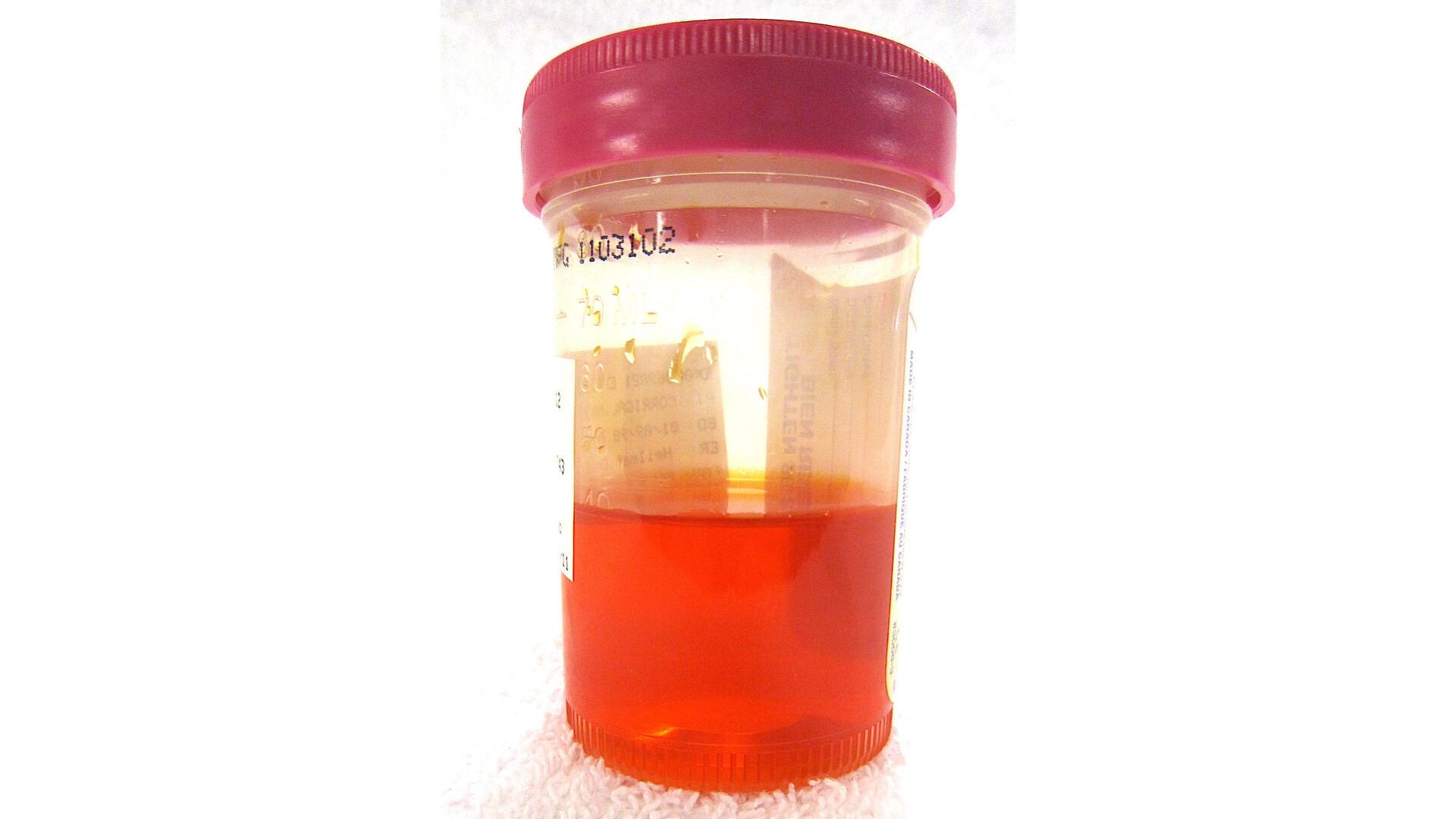
- Often feels like: one-sided flank pain that can be severe, nausea or vomiting, sometimes blood in the urine.
- What imaging may show: hydronephrosis, sometimes the stone itself, and sometimes swelling around the kidney.
- Why the report may not name it: the right test matters. Non-contrast CT is often the most sensitive study for ureteral stones, while ultrasound can miss ureteral stones and contrast on CT can make small stones harder to see in some situations.
2) Pregnancy
During pregnancy, hormonal changes relax the smooth muscle in the urinary tract, and the growing uterus can compress the ureters. This can cause hydronephrosis, often more on the right side.
- Often feels like: sometimes no symptoms, sometimes discomfort or urinary frequency.
- Why it matters: most pregnancy-related hydronephrosis is physiologic (expected), but clinicians remain alert for stones or infection because they can look similar based on symptoms alone.
3) Vesicoureteral reflux (reflux)
Reflux means urine flows backward from the bladder up toward the ureters and kidneys. It is more commonly diagnosed in childhood but can persist into adulthood or be found during workups for recurrent urinary tract infections.
- Often associated with: recurrent UTIs, kidney scarring in longstanding cases.
- How it relates to hydronephrosis: reflux can cause dilation, especially when the bladder is full or during voiding.
4) Ureteropelvic junction (UPJ) obstruction or narrowing
The ureteropelvic junction is where the kidney’s drainage system meets the ureter. Some people are born with a narrowing here, or it can be caused by scarring or compression from a nearby blood vessel.
- Often found: incidentally on imaging, or after intermittent flank pain, UTIs, or stones.
- Typical pattern: hydronephrosis in the kidney with less dilation of the ureter below the junction.
5) Bladder outlet obstruction
If urine cannot leave the bladder efficiently, pressure can back up into the ureters and kidneys, sometimes affecting both kidneys. Causes can include:
- Enlarged prostate (BPH) in men
- Urethral stricture (narrowing from scar tissue)
- Neurologic bladder dysfunction (sometimes from diabetes, spinal cord issues, certain medications, or other neurologic conditions)
- Pelvic organ prolapse in some women
This is one reason bilateral hydronephrosis often prompts clinicians to ask about urinary symptoms such as weak stream, straining, hesitancy, incomplete emptying, or new incontinence.
Other possible causes
- Tumors or masses that compress or block the ureter (in the urinary tract or nearby organs)
- Blood clots or inflammatory debris obstructing flow
- Postsurgical or radiation scarring that narrows the ureter
- Severe constipation in some cases, especially in children, can contribute to urinary retention or compression
Why the report may not name the cause
Radiologists are experts at describing what they see, but hydronephrosis is sometimes like seeing a backed-up sink without seeing the clog.
Here are common reasons a report might state “hydronephrosis” without a definitive cause:
- The blockage is intermittent: the ureter may spasm, or a stone may move, so the “why” is not captured in that moment.
- The cause is too small to visualize: tiny stones, early strictures, or subtle lesions can be missed on certain studies.
- The study was not tailored for the urinary tract: many CT scans are performed for broader abdominal reasons, and the urinary system is not the primary target.
- There may be transient, non-obstructive dilation: pregnancy is a classic example, and mild pelviectasis can be seen with an overdistended bladder that resolves after voiding.
- Symptoms and labs matter: imaging cannot tell whether you are feverish, dehydrated, or in kidney failure, but your clinician needs that context to interpret significance.
What happens next
What you need depends on your symptoms, your kidney function, and what the scan suggests. In clinic, I usually think in three parallel tracks: Is there obstruction? Is there infection? Is kidney function at risk?
A quick example from real life: I have seen patients with “moderate hydronephrosis” on an incidental CT who feel fine, but their creatinine is climbing and their bladder is not emptying. I have also seen the opposite: dramatic pain from a stone with only mild dilation. Context is everything.
Common next steps your clinician may recommend
- Urinalysis and urine culture: looks for blood, infection, and other clues.
- Blood tests: especially creatinine and estimated GFR to assess kidney function, and sometimes a complete blood count if infection is possible.
- Repeat or targeted imaging: a renal ultrasound, CT focused on stones, or other studies depending on the suspected cause.
- Bladder evaluation: if retention is suspected, measuring post-void residual urine or placing a catheter in urgent cases.
- Referral: urology is commonly involved when obstruction is suspected, hydronephrosis is moderate to severe, symptoms are significant, or kidney function is abnormal.
Sometimes, functional tests are needed
If the question is not just “Is it dilated?” but “Is it truly obstructed, and how well is the kidney working?”, clinicians may order more specialized tests, such as a diuretic renogram (often called a MAG3 scan), a CT urogram, or an MR urogram. Not everyone needs these, but they can be helpful for partial or intermittent obstruction, UPJ obstruction, or complex anatomy.
Treatment depends on the cause
Hydronephrosis itself is not the “target” of treatment. The goal is to fix what is creating the backup, for example:
- Stone management: pain control, hydration guidance, medications in select cases, and procedures if the stone will not pass or if there is infection or kidney risk.
- Relieving obstruction: a ureteral stent or nephrostomy tube may be used to drain the kidney in urgent situations.
- Addressing bladder outlet problems: medications, catheterization, or procedures depending on the cause.
- Managing reflux or UPJ obstruction: monitoring versus surgery depends on severity, infections, symptoms, and kidney function.
When it is an emergency
Some scenarios need same-day evaluation, often in an emergency department. Seek urgent care if you have hydronephrosis on imaging and any of the following:
- Fever (38°C / 100.4°F or higher), chills, or you feel very ill
- Severe flank or abdominal pain that is not controlled
- Vomiting and inability to keep fluids down
- Decreased urination or inability to urinate
- Known pregnancy with significant pain, fever, or concerning urinary symptoms
- One kidney (for example, you donated a kidney or have a solitary kidney) and there is hydronephrosis or reduced urine output
- Abnormal kidney function blood tests or rapid worsening symptoms
If obstruction and infection are present together, clinicians move quickly because the kidney needs drainage and the infection needs treatment. Importantly, fever with suspected obstruction is an emergency even if the pain is not severe. Older adults and immunocompromised patients can also present in less typical ways, so a lower threshold for urgent evaluation is reasonable.
Questions to ask
Bring your report if you can, and consider asking:
- Is the hydronephrosis mild, moderate, or severe?
- Is it unilateral or bilateral?
- Did the imaging show a stone or any obvious blockage?
- Do I need urine and blood tests today?
- Do I need repeat imaging to be sure it resolves?
- What symptoms should make me seek urgent care?
- Should I see urology, and how soon?
FAQ
Can hydronephrosis go away on its own?
Yes, it can, especially if the cause is temporary, like a stone that passes or pregnancy-related dilation after delivery. The key is confirming there is no ongoing blockage and that kidney function is protected.
Does hydronephrosis always mean a blockage?
Not always. It often suggests slowed drainage, but mild dilation can also be seen with pregnancy, reflux, or an overdistended bladder. Your clinician uses symptoms, labs, and follow-up to sort this out.
Is mild hydronephrosis serious?
Sometimes it is harmless and sometimes it is an early warning sign. Mild hydronephrosis still deserves context: your symptoms, whether it is new, and whether it persists on follow-up.
What is the difference between hydronephrosis and a kidney infection?
Hydronephrosis is a drainage finding. A kidney infection (pyelonephritis) is an infection. You can have one without the other, but when infection occurs behind an obstruction, it can become dangerous quickly.
A final word from clinic
Hydronephrosis on a report is your body’s way of waving a flag that urine flow may be disrupted. The good news is that many causes are treatable, and early attention helps protect kidney function. If you have the report in hand, your next best step is to review it with the clinician who ordered the imaging so you can connect the finding to your symptoms, labs, and a clear plan.