When I explain ureteropelvic junction obstruction to patients, I usually start with a simple picture: your kidney is like a funnel that collects urine, and your ureter is the tube that carries it down to the bladder. The spot where that funnel narrows into the tube is called the ureteropelvic junction, or UPJ. If that junction is too tight or gets kinked, urine has trouble draining. Pressure builds, the kidney looks swollen on imaging, and symptoms can range from none at all to intense pain.
UPJ obstruction can show up in infants (often discovered on prenatal ultrasound) and in adults (sometimes after years of silent backup). The key is figuring out whether the kidney is truly blocked or simply looks “full” for other reasons. That is where a diuretic renal scan becomes so helpful.
UPJ obstruction, in plain language
Ureteropelvic junction obstruction means there is a partial or complete blockage where urine exits the kidney’s collecting system (the renal pelvis) and enters the ureter.
When drainage is slowed, urine backs up and stretches the renal pelvis and calyces. On imaging reports, you may see terms like:
- Hydronephrosis (swelling of the kidney due to urine backup)
- Renal pelvic dilatation (a widened collecting system)
- Delayed drainage (urine is not clearing normally)
Not all hydronephrosis is dangerous, and not all hydronephrosis means UPJ obstruction. The job of your care team is to determine whether the dilation is causing harm, and whether fixing the narrowing will protect kidney function.
Why it happens in babies vs adults
In infants and children
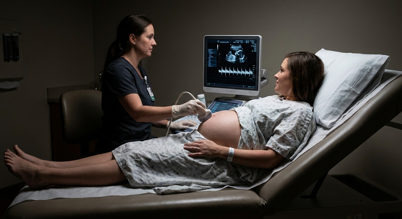
Many cases are congenital, meaning the narrowing is present from birth. It is commonly detected on prenatal ultrasound or early-life imaging done for hydronephrosis.
Common reasons include:
- A segment of the UPJ that did not form with normal muscle and elasticity
- A UPJ that is narrow, stiff, or shaped in a way that resists flow
- Less commonly, a blood vessel crossing near the UPJ that contributes to kinking as a child grows
Some mild or moderate cases can improve as a child grows, but how likely that is depends on severity and whether drainage and kidney function stay reassuring on follow-up.
In adults
Adults can have a congenital narrowing that stayed quiet for years and becomes noticeable later, often when something increases urine flow or changes anatomy. UPJ obstruction in adults can also be related to:
- Crossing blood vessels that compress the UPJ, especially with certain body positions or activity
- Less commonly, scar tissue near the UPJ, for example after prior surgery, infection, or stone-related inflammation
- Kidney stones or prior procedures that contribute to narrowing in that region
If you are an adult with new hydronephrosis, clinicians also think broadly about other causes like stones lower in the ureter, strictures, reflux, tumors, or bladder outlet obstruction. UPJ obstruction is one important possibility, but not the only one.

Symptoms and the typical pain pattern
UPJ obstruction can be tricky because symptoms are not always proportional to how dramatic the imaging looks. Some people have significant hydronephrosis and feel fine. Others have intermittent blockage that causes sharp, memorable episodes.
Adults: the classic pattern
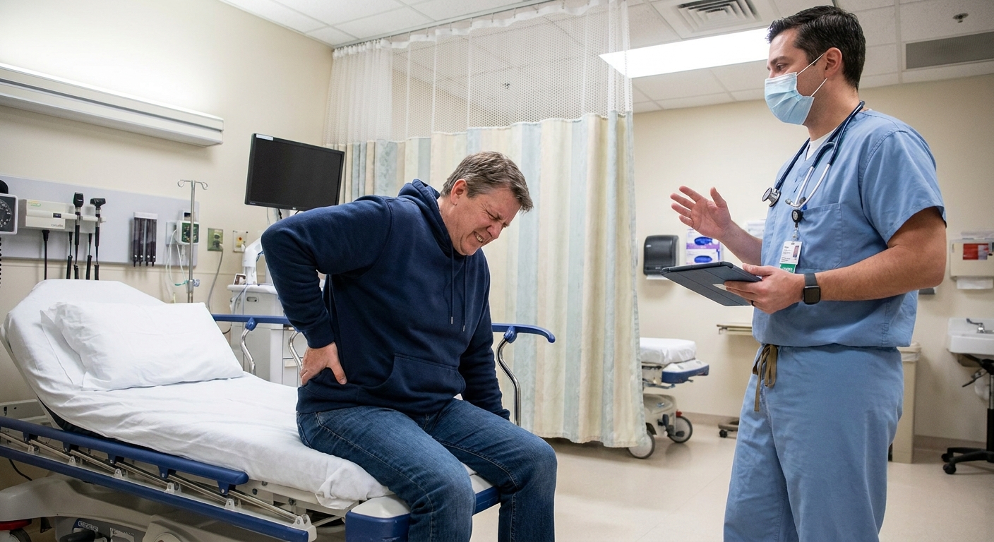
The most common symptom adults describe is flank pain on the affected side. The pain is often:
- High in the back or side, under the ribs
- Intermittent, coming in waves
- Triggered by increased urine production, such as after drinking a lot of fluid, alcohol, or caffeine, or after receiving IV fluids
- Sometimes associated with nausea or vomiting when pressure builds quickly
That “pressure surge” pattern is a clue. When the kidney makes more urine than can squeeze through the tight junction, the collecting system stretches, and stretching hurts.
Infants and children: different clues
Babies cannot tell you they have flank pain, so we look for indirect signs. Many infants with UPJ obstruction are diagnosed from imaging alone and appear well. When symptoms do occur, they can include:
- Fevers, especially if a urinary tract infection develops
- Poor feeding or irritability
- Vomiting
- Failure to thrive in more severe cases
- Abdominal mass (rare, usually with significant swelling)
In older children, symptoms can look more like adults, including flank or abdominal pain that comes and goes, sometimes during activity or after high fluid intake.
Other possible signs at any age
- Urinary tract infections
- Blood in the urine (visible or microscopic)
- Kidney stones (urine stasis can contribute)
How doctors evaluate it
Evaluation usually combines symptoms, physical exam, labs, and imaging. The goal is to answer a few practical questions:
- Is urine drainage actually impaired at the UPJ?
- Is the affected kidney losing function?
- Is this stable, or getting worse over time?
- Is there another cause of hydronephrosis that needs different treatment?
Common tests
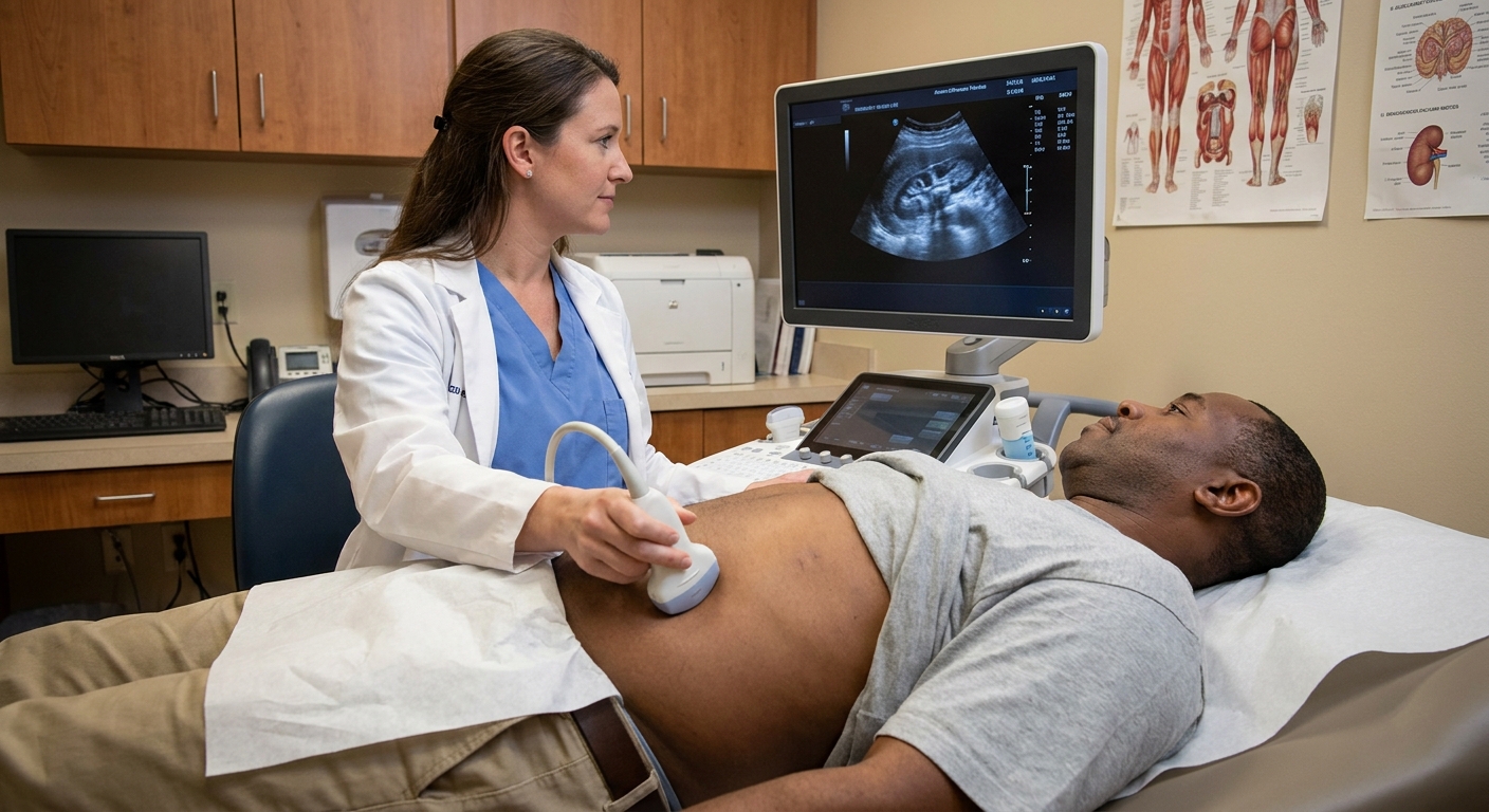
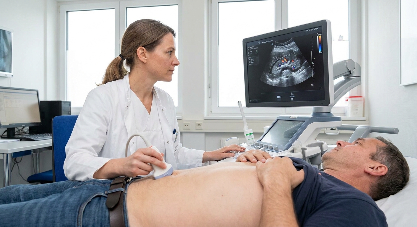
- Ultrasound: often the starting point, especially in pregnancy, infants, and follow-up monitoring
- Urinalysis and urine culture: to look for infection or blood
- Blood tests (like creatinine): to assess overall kidney function, especially if both kidneys are affected or there is concern for reduced function
- CT urography or MRI urography: sometimes used in adults to define anatomy and look for stones or a crossing vessel
The diuretic renal scan
If you remember one concept from this article, make it this: a dilated kidney is not always a blocked kidney. Sometimes the collecting system is roomy but drains adequately. Other times, the kidney is truly obstructed and pressure is building.
A diuretic renal scan is designed to sort that out. You may hear it called:
- Diuretic renography
- Lasix renal scan (Lasix is a brand name for the diuretic furosemide)
- MAG3 scan (because MAG3 is commonly used as the tracer, although some centers use other tracers)
What the test measures
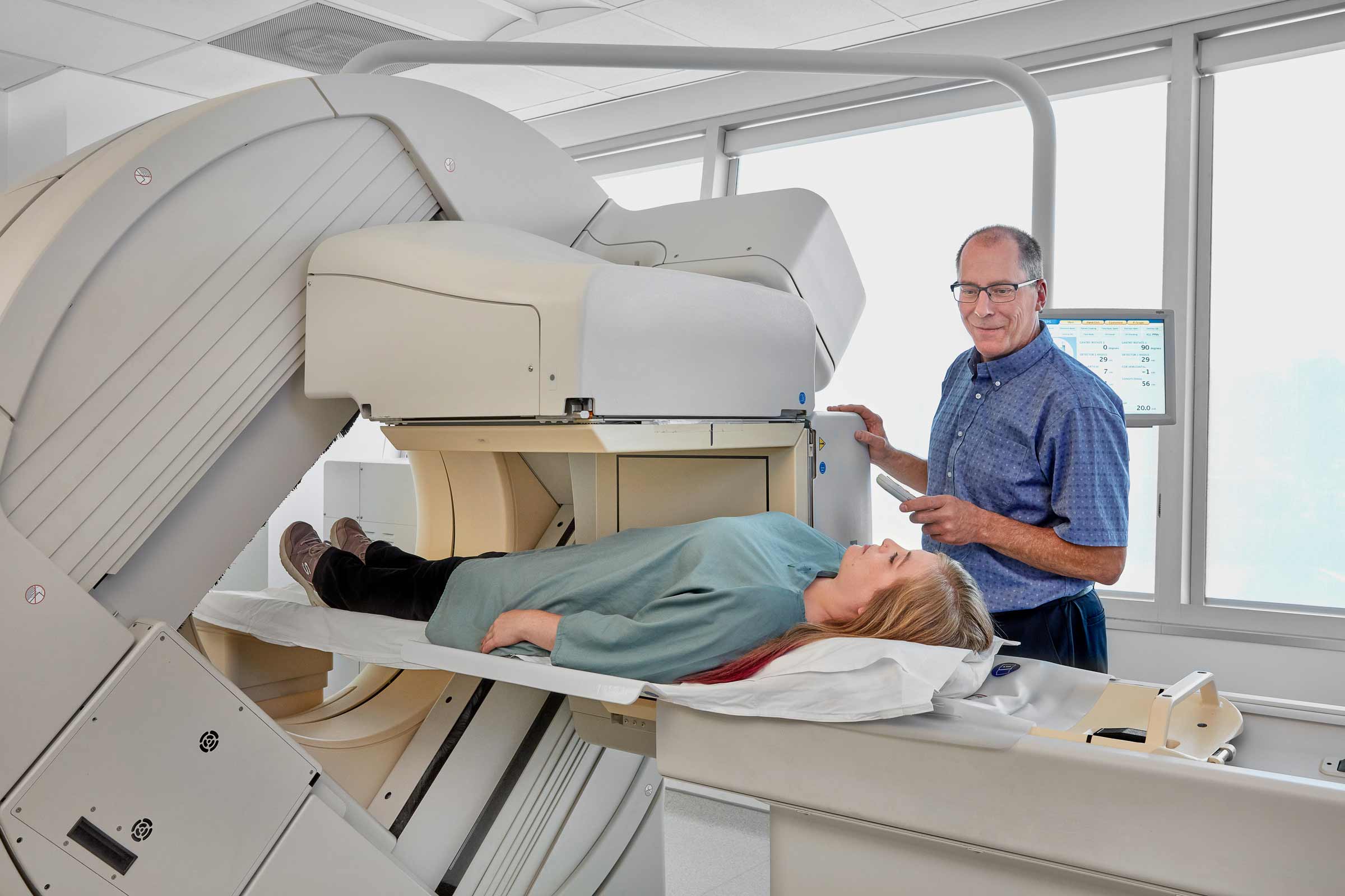
During the scan, a small amount of tracer is injected into a vein. The tracer behaves in a way that allows the camera to watch how each kidney:
- Takes up the tracer (a proxy for function and blood flow)
- Moves it into the collecting system
- Drains it down the ureter
Partway through, a diuretic is given to prompt the kidney to make more urine. This is the stress test portion.
What to expect on scan day
- Timing: many studies take roughly 45 to 90 minutes, depending on the protocol and how quickly things drain.
- Hydration: you may be asked to arrive well-hydrated or you may receive IV fluids, since dehydration can make drainage look worse than it is.
- Bladder emptying: you will usually be asked to empty your bladder during or right after the study. A very full bladder can slow drainage and affect interpretation. In select pediatric cases, a catheter may be used to keep the bladder empty so the results are easier to interpret.
- How the diuretic feels: furosemide commonly causes a strong urge to urinate for a short time. Some people feel briefly lightheaded, especially if they are not well-hydrated.
How it separates obstructed vs non-obstructed drainage
Think of it like turning on a faucet to test a drain:
- If the collecting system is simply roomy, the diuretic increases flow and the kidney washes out the tracer in a reasonable amount of time.
- If the UPJ is functionally obstructed, the extra flow cannot get through and tracer lingers in the kidney, showing delayed washout.
The scan report may mention concepts like drainage half-time (how long it takes for activity to drop by half after the diuretic) and differential renal function (how much each kidney contributes to total kidney function). These details help urologists decide whether observation is safe or whether the kidney is at risk.
One important nuance: delayed washout does not always mean a fixed, surgical obstruction. Results can be affected by hydration status, bladder fullness or retention, very poor kidney function, or a very capacious collecting system (for example an extrarenal pelvis). That is why your clinician interprets the scan in context, not as a standalone number.
If your results are confusing, it is reasonable to ask, “Does this look like true obstruction, and is my kidney function being affected?”
When pyeloplasty comes up
Pyeloplasty is the most common surgical repair for UPJ obstruction. In simple terms, the surgeon removes or bypasses the narrowed segment and reconnects the healthy portions to create a wider, better-draining junction.
Not everyone with UPJ obstruction needs surgery. Many infants are monitored because some cases improve with growth, especially when kidney function is preserved and follow-up imaging stays stable. Many adults with minimal symptoms and preserved function can also be safely observed.
Why surgeons consider repair
While exact thresholds vary by age, anatomy, and surgeon preference, pyeloplasty is more likely to be discussed when one or more of the following is present:
- Decreasing function of the affected kidney over time on renal scan
- Poor drainage consistent with functional obstruction on a diuretic renal scan
- Recurrent or severe flank pain that fits an obstruction pattern
- Recurrent urinary tract infections, especially febrile UTIs
- Complications such as stones related to urine stasis
- Worsening hydronephrosis on follow-up imaging
Other treatments you may hear about
Pyeloplasty is the mainstay, but there are other options in selected situations:
- Temporary ureteral stent or nephrostomy tube: used to relieve pressure and protect the kidney in acute situations (for example severe pain with significant obstruction, or obstruction with infection) or as a bridge to definitive repair.
- Endopyelotomy: an internal incision of the narrowed segment performed endoscopically in selected adults. It can be appropriate in specific anatomy, but success rates can be lower than pyeloplasty for certain causes (such as a crossing vessel).
Follow-up and monitoring
If observation is chosen, follow-up often includes serial ultrasounds and symptom check-ins. A repeat diuretic renal scan may be used if there is a question about drainage over time or whether kidney function is changing.
Recovery details and what to expect after surgery matter a lot, but they deserve their own focused discussion. If pyeloplasty has been recommended to you or your child, ask for a clear explanation of the goal of surgery, what test results the team is responding to, and how success will be measured afterward.
When to seek urgent care
Most UPJ obstruction evaluations happen in a calm outpatient setting, but a few symptoms should prompt urgent assessment, especially if you have known hydronephrosis:
- Fever with chills, especially with urinary symptoms (could signal a kidney infection)
- Severe flank pain that does not improve, especially with vomiting or inability to keep fluids down
- Markedly decreased urine output, especially if you have one kidney, known problems on both sides, or significant dehydration
- Visible blood in the urine with pain or clots
If an obstructed system becomes infected, that is an emergency. Trust your instincts and get help quickly.
Questions to ask
- Does my imaging suggest UPJ obstruction, or could this be another cause of hydronephrosis?
- What did the diuretic renal scan show about drainage and split kidney function?
- Is my kidney function stable over time?
- What symptoms should prompt me to call you sooner?
- If we are observing, what is the follow-up plan and what change would trigger surgery?
If you are reading an imaging report and feeling overwhelmed, you are not alone. The words can sound alarming. The good news is that UPJ obstruction is a well-studied condition, and modern imaging gives clinicians a practical way to separate a kidney that is truly obstructed from one that is simply dilated but draining.