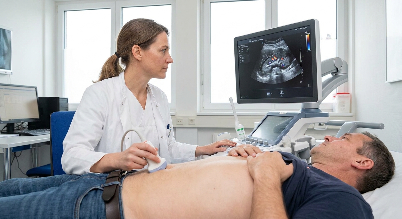
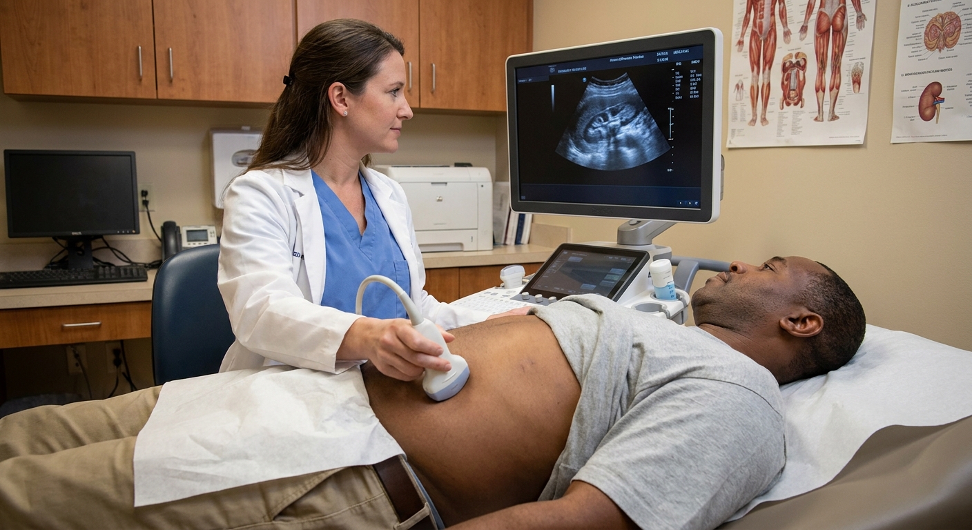
Getting a call that something looked “dilated” or “swollen” on your baby’s anatomy scan can make your stomach drop. I have sat with many families in that exact moment, and the first thing I want you to know is this: fetal hydronephrosis is common, and in many pregnancies it is mild and temporary.
On a prenatal ultrasound, “hydronephrosis” usually refers to extra fluid in the part of the kidney that collects urine. The ultrasound is not seeing pain, kidney failure, or a stone. It is seeing a measurement that is a little larger than expected for that stage of pregnancy.
How common? Depending on how a practice defines and measures it, urinary tract dilation is often reported in roughly 1% to 5% of pregnancies.
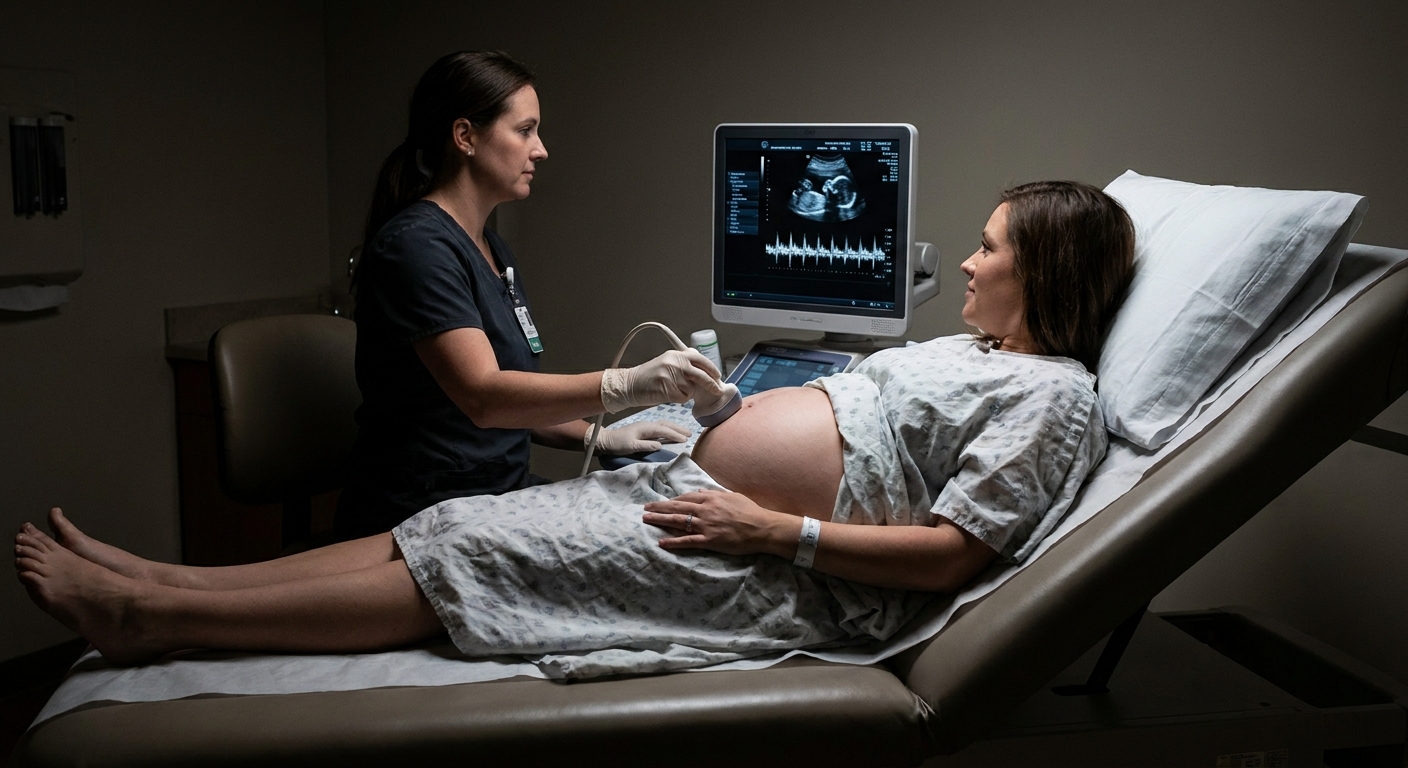
This page stays focused on what hydronephrosis means in a fetus and newborn, which is different from the wording you might see on an adult imaging report.
What it means
Babies begin making urine early in pregnancy. That urine drains from each kidney down a tube called a ureter into the bladder. Later in pregnancy, fetal urination becomes a major contributor to amniotic fluid volume.
On ultrasound, the radiologist or maternal-fetal medicine specialist is usually looking at the renal pelvis, the central “funnel” area where urine collects before draining.
Fetal hydronephrosis (also called antenatal hydronephrosis or urinary tract dilation, often shortened to UTD) means that this collecting area looks wider than expected.
Hydronephrosis can involve:
- One kidney (unilateral), which is commonly seen
- Both kidneys (bilateral), which often prompts closer monitoring
- Additional dilation of the ureter, bladder changes, or both
How often it goes away
UTD is among the most common findings on the mid-pregnancy anatomy scan. In day-to-day clinic language, it is often described as “mild kidney swelling” or “fluid in the kidney.”
The reassuring part is that mild dilation frequently improves on its own during pregnancy or after birth. A common reason is simply that the baby’s urinary system is still developing, and drainage can look a little roomy before everything matures.
Even when it does not fully resolve before delivery, many babies with mild findings do well with monitoring only and never need surgery.
The measurement that matters
When families ask me, “How worried should I be?”, my next question is almost always: What was the measurement?
The key number on most reports is the anteroposterior renal pelvic diameter (often shortened to APRPD or “renal pelvis measurement”). This is the width of the fluid space in the kidney on a specific ultrasound view.
Important note: Cutoffs vary by guideline and by hospital. Some reports use the UTD classification (UTD A1 vs UTD A2 to A3 prenatally) instead of the words “mild, moderate, severe.” Your team will apply the system they use locally.
Typical ranges
As a general, commonly used framework based mainly on the APRPD measurement:
- Second trimester (about 16 to 27 weeks): mild around 4 to 7 mm, often called moderate if 7 to 10 mm, and more concerning when 10 mm or more or when other findings are present
- Third trimester (28 weeks and later): mild around 7 to 9 mm, often called moderate if 9 to 15 mm, and more concerning when 15 mm or more or when other findings are present
In plain language, “mild” usually means the kidney looks a little roomy but the rest of the urinary tract looks reassuring. “Moderate” and “severe” generally mean the dilation is larger, more persistent, or paired with other clues that there may be a drainage issue that needs closer follow-up.
If you have your ultrasound report, you can ask your OB team: “What was the renal pelvis measurement in millimeters, was it one kidney or both, and is this UTD A1 or UTD A2 to A3 in your system?” That combination often brings clarity.
Other findings that matter
Hydronephrosis is not a one-size-fits-all finding. The same kidney measurement can mean different things depending on what else is seen on the scan.
One kidney vs both
- Unilateral UTD often has a more straightforward course, because the other kidney is usually working normally.
- Bilateral UTD does not automatically mean something severe, but it more often leads to closer prenatal follow-up and a clearer postnatal plan.
The bladder
Your clinician may comment on whether the bladder:
- Fills and empties normally during the scan
- Looks unusually large or thick-walled
- Shows other clues that urine flow could be obstructed
A bladder that is persistently very enlarged or does not appear to empty can raise concern for a lower urinary tract issue and typically triggers maternal-fetal medicine involvement and more frequent monitoring.
The ureters
If the ureter looks dilated (sometimes called hydroureter), it can suggest that urine is backing up beyond the kidney, which can change postnatal testing recommendations.
Amniotic fluid
This is a big one. Baby urine is a major contributor to amniotic fluid in the second half of pregnancy.
- Normal amniotic fluid is generally reassuring.
- Low amniotic fluid (often called oligohydramnios) can signal that the baby is not making or releasing urine normally and needs prompt, specialized evaluation.
Kidney tissue
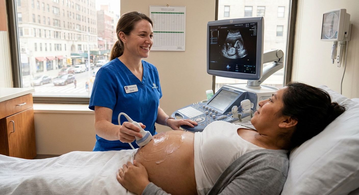
Beyond the “fluid space” measurement, the ultrasound may comment on the kidney’s appearance: whether the kidney tissue looks normal, whether there are cysts, or whether the collecting system has a more complex appearance. Those details help determine whether this is likely a simple, temporary dilation or something that needs closer surveillance.
Common causes
In plain language, fetal hydronephrosis usually comes down to one of two categories:
- Urine is draining a bit slowly at some point along the pathway
- Urine is flowing backward toward the kidney
Some common underlying causes your care team may mention include:
- Transient dilation: the most common explanation for mild cases, and it often resolves without intervention
- Ureteropelvic junction (UPJ) narrowing: a tight spot where the kidney connects to the ureter
- Vesicoureteral reflux (VUR): urine refluxing from the bladder back up toward the kidney
- Ureterovesical junction (UVJ) narrowing: a tight spot where the ureter connects to the bladder
- Posterior urethral valves: a condition affecting the urethra in male fetuses that can obstruct urine outflow and typically requires prompt evaluation and coordinated planning with specialists
It can feel overwhelming to hear a list like this. The important point is that the ultrasound pattern (measurement plus the bladder, ureters, kidney appearance, and amniotic fluid) helps your team narrow what is most likely.
Prenatal follow-up
Most families will be offered a follow-up ultrasound, often in the third trimester, to see if the dilation is improving, stable, or increasing.
Depending on the findings, your OB may:
- Repeat an ultrasound in several weeks
- Refer you to maternal-fetal medicine for a more detailed look
- Arrange a prenatal consult with pediatric urology so you already have a plan in place for after birth
In many mild, isolated cases, the plan is simply: monitor during pregnancy and check again after delivery.
One gentle reminder: there is usually nothing you need to change day to day based on this ultrasound finding alone. Do not restrict your fluids or try to “treat” it with diet changes unless your own clinician has given you specific instructions for a different reason.
Prognosis by severity
If you want the big-picture expectations, I usually frame it like this:
- Mild, isolated UTD: most commonly improves or resolves, either before birth or in early infancy, and often only needs follow-up ultrasound.
- Moderate UTD: many cases still improve, but the chance of needing additional imaging, specialist follow-up, or longer-term monitoring is higher.
- Severe UTD or UTD with other findings (dilated ureter, abnormal bladder, low amniotic fluid): needs closer surveillance because it has a higher chance of representing an obstruction or reflux that could affect kidney health without monitoring.
Genetic screening question
Some parents notice that mild renal pelvic dilation is sometimes discussed as a “soft marker” on ultrasound. When UTD is isolated and prior genetic screening is reassuring, it often does not substantially change risk. Still, it is reasonable to ask your OB or MFM team whether your ultrasound finding changes any recommendations for reviewing or offering genetic screening in your specific pregnancy.
After birth: ultrasound timing
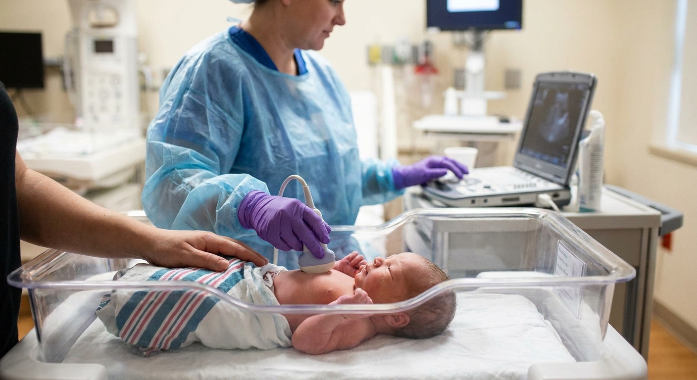
One detail that surprises a lot of parents is that the first kidney ultrasound is often not done immediately after birth unless there are higher-risk findings.
Newborns are naturally a bit dehydrated right after delivery, especially in the first 24 to 48 hours, which can make dilation look smaller than it really is. For that reason, many clinicians prefer a renal and bladder ultrasound:
- After 48 hours of life for many babies, and often
- Within the first 1 to 2 weeks for lower-risk cases (depending on local protocol)
High-risk situations may be imaged sooner or managed more urgently, including severe bilateral dilation, concerning bladder findings, suspected outlet obstruction, or low amniotic fluid. Your team will choose timing that matches your baby’s risk level and the hospital’s protocol.
What the newborn ultrasound checks
- Whether dilation is still present
- Whether one or both kidneys are involved
- Whether the ureters look enlarged
- Bladder wall and bladder emptying pattern
Many babies with mild prenatal dilation have a normal or improved ultrasound after birth.
When more tests are needed
If the postnatal ultrasound shows moderate to severe dilation, ureter dilation, abnormal bladder findings, or other red flags, your pediatrician may recommend additional testing. Two commonly discussed tests are:
- VCUG (voiding cystourethrogram): evaluates for vesicoureteral reflux and also looks at the urethra and bladder outlet anatomy, which matters when conditions like posterior urethral valves are a concern
- Diuretic renal scan (such as MAG3 in some settings): helps assess drainage and how well each kidney is functioning, usually ordered by specialists
Not every baby needs these tests. In fact, avoiding unnecessary testing is part of good care. The decision is typically driven by how the kidneys, ureters, and bladder look on ultrasound and whether the baby has urinary symptoms or urinary tract infections.
When pediatric urology is involved
Some babies are referred to pediatric urology prenatally, but many are referred after the first newborn ultrasound.
Referral is more likely when:
- Dilation is moderate to severe
- Both kidneys are affected
- The ureter is dilated
- The bladder looks abnormal or there are concerns for outlet obstruction
- Amniotic fluid was low
- Dilation persists or worsens on postnatal imaging
Pediatric urologists focus on protecting kidney function and preventing complications. A lot of visits are about monitoring growth and repeat imaging, not rushing to procedures.
Antibiotics
Parents often hear about “preventive antibiotics” and understandably worry. In some situations, clinicians prescribe a low daily antibiotic to reduce the risk of urinary tract infection, especially when there is higher-grade dilation, ureter dilation, known reflux, or other risk factors.
In mild, isolated cases, preventive antibiotics are often not needed. This is a personalized decision, so ask your baby’s clinician to explain the “why” in your specific situation.
What to watch for
Many newborns with prenatal hydronephrosis look completely well. Still, it helps to know what should prompt a call to your pediatrician urgently, especially before the follow-up imaging is completed:
- Fever in an infant under 3 months (this is always urgent)
- Poor feeding, unusual sleepiness, or vomiting
- Fewer wet diapers than expected
- Foul-smelling urine (hard to judge in newborns, but mention it if you notice it)
If a young infant develops a fever, clinicians often evaluate for a urinary tract infection along with other causes.
Questions to ask
When you are anxious, it is hard to remember what you wanted to ask. These questions tend to get families the most useful, concrete answers:
- What was the kidney measurement in millimeters, and which side is affected?
- Is this considered mild, moderate, or severe in our practice, or is it classified as UTD A1 vs A2 to A3?
- Are both kidneys involved?
- Did the report mention the ureters or the bladder?
- Is amniotic fluid normal?
- When do you want the next prenatal ultrasound?
- What is the plan for the newborn renal and bladder ultrasound, and who orders it?
- At what point do you refer to pediatric urology?
Bottom line
Fetal hydronephrosis on a prenatal ultrasound usually means the urine-collecting area of one or both kidneys measures a bit larger than expected. Mild cases often resolve or improve significantly, and many babies only need follow-up imaging.
The plan is shaped by a few key details: the measurement in millimeters, whether it is one kidney or both, what the bladder and ureters look like, and whether amniotic fluid is normal. After birth, most babies have a renal and bladder ultrasound on a timeline chosen to give accurate information, and pediatric urology is brought in when the findings suggest closer monitoring or additional testing.
If you are sitting with this news right now, take a breath. Ask for the measurement, ask what else was seen, and ask for the post-birth plan. Clear information is one of the best anxiety reducers we have.
FAQ
Is fetal hydronephrosis the same as a kidney infection?
No. Hydronephrosis is an ultrasound finding describing dilation in the urine-collecting system. It is not a diagnosis of infection. Infections are diagnosed after birth using symptoms and urine testing.
Does fetal hydronephrosis mean my baby will have kidney damage?
Not necessarily. Many mild cases do not affect kidney function. The purpose of follow-up ultrasounds is to identify the smaller group of babies who need closer monitoring to protect kidney health.
Can fetal hydronephrosis affect delivery plans?
Most of the time, no. Delivery decisions are usually based on overall pregnancy health. In higher-risk urinary findings, your OB and specialists may coordinate where the baby should be delivered to ensure timely newborn evaluation.