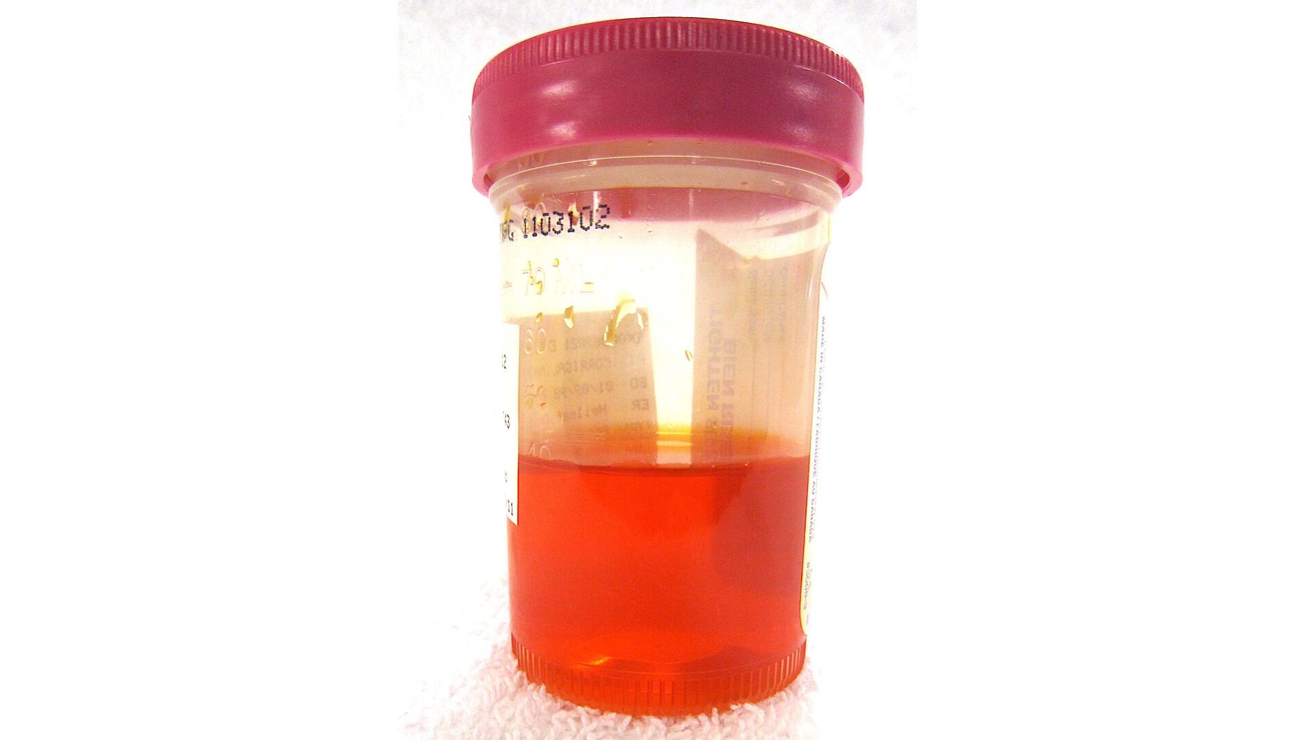
When a child has a urinary tract infection (UTI), especially one that comes with fever, it can feel like the finish line is finishing antibiotics and seeing symptoms resolve. Then the pediatrician says, “Let’s schedule a kidney ultrasound.”
I have cared for many families who hear that and immediately think something must be seriously wrong. Most of the time, that is not the case. A kidney and bladder ultrasound is often a careful, preventive next step to make sure your child’s urinary system is built and draining in a way that makes future infections less likely and less harmful.

Below I will walk you through why imaging is sometimes recommended after a UTI, what common findings like hydronephrosis can mean, what a normal report actually reassures us about, and which children may need additional testing for reflux.
What doctors are trying to answer
In pediatrics, the main reason to image after a UTI is not to “hunt for problems,” but to reduce the chance of missed anatomy or drainage patterns that could set a child up for repeat infections or kidney stress.
A kidney and bladder ultrasound, sometimes written as RBUS (renal bladder ultrasound), helps your child’s clinician answer a few practical questions:
- Are both kidneys present and about the expected size?
- Is urine draining well from the kidneys down to the bladder? We look for dilation that suggests backup.
- Is the bladder emptying normally? A very full bladder after voiding can raise UTI risk.
- Are there structural differences that might increase infection risk, such as dilation of the urinary tract or an obstruction?
Ultrasound is popular for this job because it does not use radiation, it is noninvasive, and it can provide a lot of information without needles or sedation in most children.
When ultrasound is recommended
Clinicians do not image every child after every bladder infection. Imaging decisions depend on age, whether there was fever (suggesting the kidneys may have been involved), how sick the child was, and whether there are repeat infections. Guidelines also vary by country, health system, and specialist group.
Common situations where ultrasound is often ordered
- Infants and young children after a first febrile UTI. Many practices are especially likely to recommend RBUS in children under 2 years old (often discussed as ages 2 to 24 months), because anatomy issues are more likely to show up early and can change management.
- Any age with recurrent UTIs. Repeating infections raises the odds of an underlying drainage issue or a bladder-emptying problem.
- UTI with an atypical course. Examples include poor response to antibiotics within the expected window, severe illness, unusual bacteria, or concerns for obstruction.
- History that suggests urinary tract differences. Prenatal ultrasound concerns, known kidney issues, poor growth, high blood pressure, or abnormal urine findings can influence the decision.
Your child’s pediatrician or pediatric urologist may also time the ultrasound based on the clinical situation. Often, the ultrasound is scheduled after the child is improving, but it may be done sooner if there is concern for blockage, complications, or a child who remains quite ill.
If you are unsure why imaging was recommended for your child specifically, ask one direct question: “What decision will this ultrasound help you make?” A good clinician should be able to answer that clearly.
What the ultrasound is like
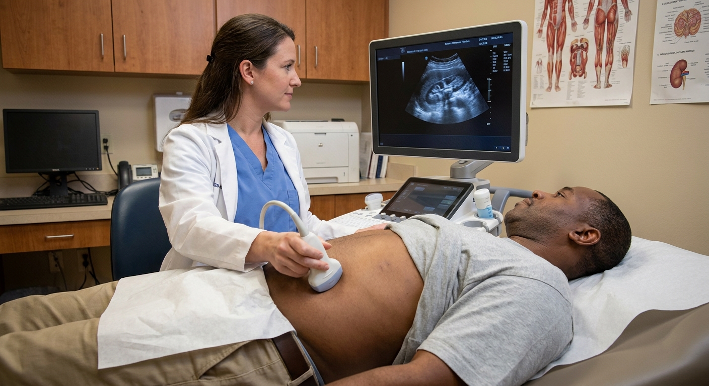
An ultrasound is usually straightforward. A technologist places warm gel on the skin and uses a handheld probe to take images of the kidneys and bladder. The test is painless, though some kids dislike the cold gel or the pressure of the probe.
Parents often ask about preparation. Policies vary, but many facilities want the child to arrive with a comfortably full bladder, because it can help evaluate the bladder wall and how well the bladder empties.

How to read common results
Ultrasound reports can sound intimidating because they are written for medical teams, not for families. Here is how I translate the most common terms parents see after a UTI.
“Normal renal ultrasound” or “no hydronephrosis”
A normal study is reassuring. It suggests:
- The kidneys look structurally typical and are not obviously swollen from backed-up urine.
- There is no clear ultrasound evidence of an obstruction.
- The bladder appearance is not raising red flags at the moment of the scan.
Important nuance: a normal ultrasound does not rule out every possible issue. In particular, it cannot reliably rule out vesicoureteral reflux (reflux of urine from the bladder back toward the kidneys). That is tested with a different study.
Hydronephrosis
Hydronephrosis means the part of the kidney that collects urine looks stretched or swollen. Families often hear “swollen kidney,” and that phrase can be scary. Hydronephrosis is a description, not a diagnosis by itself.
It can show up for several reasons, including:
- Drainage that is slower than expected somewhere along the pathway.
- Vesicoureteral reflux, where urine can travel backward under pressure.
- An anatomic narrowing or blockage (less common, but important to identify).
- Transient collecting-system dilation that can be seen around an acute illness and is sometimes influenced by hydration status or how full the bladder is during the study.
Reports often include a severity estimate such as mild, moderate, or severe. In general, the more significant the hydronephrosis, the more likely your child’s clinician will recommend follow-up imaging, specialist referral, or additional testing.
Hydroureter or “ureteral dilation”
The ureter is the tube that carries urine from the kidney to the bladder. If it looks widened, it may suggest reflux or a downstream obstruction. This finding often prompts a closer look at the overall clinical story, including how many UTIs your child has had and whether they were febrile.
“Renal scarring” or concerns about scarring
This is one of the most anxiety-provoking phrases for parents, and it deserves careful context.
Ultrasound is not the best test for confirming small kidney scars. Sometimes ultrasound can show changes that suggest prior injury, such as an irregular outline or differences in kidney tissue appearance. If scarring is truly a concern, clinicians may consider other studies depending on the situation, your child’s age, and the severity of infections.
What scarring generally means clinically is that there has likely been inflammation or infection involving kidney tissue in the past. The reason pediatric teams care is that significant scarring can, in some cases, increase risk for high blood pressure or reduced kidney function later in life. The good news is that most children with a single, promptly treated UTI do not go on to have clinically significant kidney damage.
Bladder findings: wall thickening, debris, or large post-void residual
Sometimes the report mentions bladder features such as:
- Debris in the bladder, which can be seen with infection or inflammation. It is also nonspecific and may be seen with concentrated urine or crystals.
- Bladder wall thickening, which can happen with irritation, frequent holding, or chronic voiding issues.
- Post-void residual, meaning urine remains after the child tries to empty. This can raise UTI risk and may point toward bowel and bladder dysfunction, constipation, or learned holding behaviors in older children.
These findings often lead to practical next steps that help many families, such as constipation management, timed voiding, and hydration routines.
What ultrasound cannot tell us
One reason ultrasound is a first step is that it answers some questions very well, but not all of them. Depending on the child, an RBUS may not be able to:
- Diagnose or grade reflux (vesicoureteral reflux) reliably
- Confirm or date subtle scarring with certainty
- Prove how well the kidneys function or how well urine drains over time (some questions require different imaging)
- Catch every small stone or subtle abnormality
Why reflux testing is not for every child
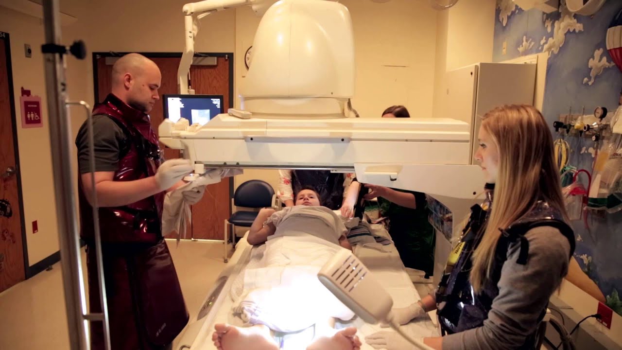
Many parents hear about a VCUG and wonder why it is not automatically ordered. A VCUG (voiding cystourethrogram) is a test that looks for vesicoureteral reflux by filling the bladder with contrast through a catheter and taking X-ray images while the child voids.
VCUG can be very helpful in specific situations, but it is more invasive than ultrasound and it uses radiation. For those reasons, many pediatric teams reserve it for children who are more likely to benefit from the answer.
Children who may need reflux testing later
Your clinician may discuss VCUG if your child has one or more of the following:
- Abnormal ultrasound findings such as moderate to severe hydronephrosis, ureteral dilation, or signs suggesting obstruction
- Recurrent febrile UTIs, especially in younger children
- UTI with an unusual or severe presentation, such as poor response to treatment or serious illness
- Concerns for bladder outlet obstruction or urethral issues, which are uncommon but important to diagnose (for example, posterior urethral valves in infant boys)
On the other hand, a child with a single, straightforward febrile UTI and a normal ultrasound may not need reflux testing right away. In that situation, many teams focus on prevention, close follow-up, and reassessment if infections recur.
What happens after the ultrasound
Next steps depend on the results and your child’s history.
If the ultrasound is normal
- Your pediatrician may simply document the result and focus on prevention: good hydration, regular bathroom breaks, and constipation control.
- If your child has another febrile UTI, imaging discussions may come up again because the risk profile changes with recurrence.
If the ultrasound shows mild hydronephrosis
- Your clinician may recommend a repeat ultrasound after a period of time to see if it resolves.
- They may also ask about voiding habits and constipation, because bladder and bowel patterns can affect urinary drainage.
If the ultrasound shows moderate to severe hydronephrosis or ureteral dilation
- Referral to pediatric urology or pediatric nephrology is common.
- Additional testing may be recommended, which could include VCUG or other imaging depending on the suspected cause.
If the report suggests scarring or kidney size differences
- Your child may need follow-up for blood pressure checks and, sometimes, lab work to monitor kidney function.
- Specialist input can help decide whether additional imaging is needed and how often to monitor over time.
Prevention basics that help
If your child’s clinician suspects bowel and bladder dysfunction, the most effective steps are often simple and consistent. Examples I commonly recommend families discuss with their pediatrician include:
- Timed voiding (for example, trying every 2 to 3 hours during the day, not just “when it hurts” or “when it is urgent”)
- Constipation treatment with a clear plan, because constipation and UTIs are frequent partners
- Hydration routines so urine is not overly concentrated
These are not one-size-fits-all, but they are a good starting point for many kids.
Questions to ask at follow-up
When you are handed an imaging report, it is easy to feel like you need to interpret every line yourself. You do not. What you need is a clear plan.
- What did you see on the ultrasound in plain language?
- Do these findings change my child’s treatment or prevention plan today?
- Do we need repeat imaging? If yes, when and why?
- Does my child need reflux testing (VCUG)? If not, what would make you recommend it later?
- Should we address constipation or voiding habits? If yes, what exact steps should we start this week?
When to call urgently
After a UTI, I want families to feel confident about what is “watch and wait” versus what needs immediate care.
Contact your child’s clinician promptly or seek urgent care if your child has:
- Fever returning after starting antibiotics or fever that does not improve as expected
- Vomiting, dehydration, or inability to keep down fluids
- New flank or back pain, or your child is very uncomfortable
- Signs of worsening illness such as unusual sleepiness, fast breathing, or poor feeding in an infant
- Blood in the urine that is visible, especially with pain
If your child is under 2 to 3 months old with any fever, that is typically an urgent situation and should be evaluated right away. When in doubt, follow your clinician’s or local emergency department guidance.
Bottom line
A kidney and bladder ultrasound after a febrile UTI is a common, thoughtful way pediatric teams check for urinary tract anatomy or drainage patterns that could raise the risk of repeat infections or kidney stress. A normal study is reassuring, hydronephrosis is a descriptive finding that often needs context and sometimes follow-up, and reflux testing is usually reserved for children with recurrent infections or abnormal ultrasound findings.
If you have your child’s ultrasound wording in front of you and want help translating it into questions for your next appointment, bring the exact phrases to the visit and ask what they mean for your child’s plan, not just what they mean on paper.
FAQ
Is a kidney ultrasound painful for a child?
No. Ultrasound is noninvasive. Most children tolerate it well, though the gel may feel cold and the probe pressure can be mildly uncomfortable.
Does hydronephrosis mean my child has permanent kidney damage?
Not necessarily. Hydronephrosis means dilation of the urine-collecting area of the kidney. It can be mild and temporary, or related to drainage issues or reflux. Your clinician will interpret it alongside severity, symptoms, and whether UTIs recur.
If the ultrasound is normal, can my child still have reflux?
Yes. Vesicoureteral reflux is not reliably ruled out by ultrasound alone. A VCUG is the classic test for reflux, but it is usually reserved for children with certain risk factors, repeat febrile UTIs, or abnormal ultrasound results.
Why not do every test at once after the first UTI?
In pediatrics we try to balance getting useful answers with minimizing invasive testing and radiation exposure. Ultrasound is a low-risk first step that helps decide whether further testing is likely to change care.