When you open a prostate biopsy report, the numbers can feel blunt and mysterious. I have sat with many patients who were less afraid of the biopsy itself than they were of the pathology line that followed. The Gleason score is one of the most important parts of that report, but it is not a “how much cancer do I have” number. It is a measure of how aggressive the cancer cells look under the microscope.
This article will walk you through what the Gleason patterns are, how the score is built, what Grade Groups 1 through 5 mean, and why 3+4 is not the same as 4+3. Then we will connect the dots between Gleason, PSA, and MRI, because real-world decisions are made using the whole picture.

Gleason score in plain language
The Gleason system grades prostate cancer based on pattern, meaning how the prostate gland tissue architecture looks compared with normal prostate tissue.
Pathologists look at the biopsy cores and identify the two most prominent patterns of cancer present:
- Primary pattern: the most common pattern seen
- Secondary pattern: the next most common pattern seen
Each pattern is assigned a number. Today, in most biopsy reports, the patterns you will see are typically 3, 4, or 5. (Patterns 1 and 2 are rarely used in modern reporting for needle biopsies.)
The Gleason score is written as primary + secondary = total, for example 3+4=7.
What Gleason patterns 3, 4, and 5 mean
You can think of Gleason patterns as a “how organized vs. chaotic” scale of the cancer’s structure.
Gleason pattern 3
Pattern 3 cells still form glands that look fairly recognizable, just more crowded and abnormal than normal prostate tissue. In many cases, this is considered lower-grade disease.
Gleason pattern 4
Pattern 4 shows more distorted gland structure. Instead of neat, separate glands, the glands may look fused, poorly formed, or cribriform (a more complex growth pattern). Pattern 4 is a key reason doctors look closely at the amount of pattern 4, along with MRI, PSA density, and biopsy volume, when deciding whether monitoring is still appropriate.
Gleason pattern 5
Pattern 5 is the least organized. The cancer may grow in sheets or single cells with minimal gland formation. This is generally considered high-grade and more likely to behave aggressively.

How the Gleason score is calculated
The key idea is that the Gleason score reflects the main patterns present in the sample, not an average, and not automatically the “worst tiny speck.” Reporting conventions vary by lab, but in general:
- If there is a small but clinically meaningful higher-grade component, some pathologists may incorporate it as the secondary pattern.
- Some reports may mention a minor or tertiary higher-grade pattern, more commonly in prostatectomy specimens than in needle biopsies.
Common totals you might see include:
- 6 (3+3)
- 7 (3+4 or 4+3) (7 is very common)
- 8 (4+4, 3+5, or 5+3) (3+5 and 5+3 are less common)
- 9 (4+5 or 5+4)
- 10 (5+5)
Even though Gleason 6 sounds “middle of the road” on a 1 to 10 scale, it is actually the lowest score commonly used on needle biopsy today. Gleason 6 (Grade Group 1) often behaves like low-grade cancer, but it is still cancer and it still deserves a thoughtful plan.
Grade Groups 1 through 5
Most modern pathology reports include a Grade Group. It is still based on Gleason patterns, but it is designed to be more intuitive.
- Grade Group 1: Gleason 3+3=6 (lowest grade commonly reported)
- Grade Group 2: Gleason 3+4=7
- Grade Group 3: Gleason 4+3=7
- Grade Group 4: Gleason 8 (4+4, 3+5, or 5+3)
- Grade Group 5: Gleason 9 to 10 (4+5, 5+4, or 5+5)
As Grade Group increases, the risk of adverse outcomes goes up. That can include faster growth, a higher chance of recurrence after treatment, and a higher chance of spread over time. Risk is never determined by Grade Group alone.
Why 3+4 is different from 4+3
This is one of the most important “small details” in a biopsy report.
Gleason 3+4=7 (Grade Group 2)
Most of what the pathologist sees is pattern 3, with a smaller component of pattern 4. Many people with 3+4 disease are still candidates for active surveillance, depending on other factors like how much cancer was found, whether cribriform or intraductal features are present, and what the MRI and PSA density suggest.
Gleason 4+3=7 (Grade Group 3)
Here, pattern 4 is the dominant pattern. That typically signals a higher risk cancer than 3+4, even though the total is still 7. In practice, 4+3 is more likely to lead to a recommendation for active treatment rather than monitoring alone, though the best plan is individualized.
If you take one thing away from this article, let it be this: the order of the numbers matters.

Other biopsy details that matter
Gleason and Grade Group tell you how the cells look. Your report often includes other clues about how much and where.
Number of positive cores
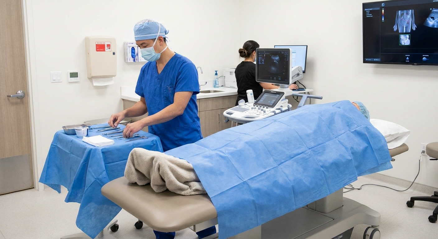
A biopsy takes multiple tissue samples called cores. Reports may say something like “3 of 12 cores positive.” In general, fewer positive cores can mean lower volume disease, but location and MRI targeting matter.
Percent involvement in each core
You may see “15 percent of the core involved” or a measurement in millimeters. This helps estimate tumor volume and can influence whether surveillance is reasonable.
Perineural invasion
This means cancer cells are seen tracking along a nerve within the prostate. It can be associated with higher-risk features in some settings, but it is not an automatic sign of spread outside the prostate. It is one piece of the puzzle.
Cribriform pattern or intraductal carcinoma
Some reports mention cribriform growth (a subtype of pattern 4) or intraductal carcinoma (often written as IDC-P). These findings can be associated with more aggressive behavior, and many specialists weigh them heavily. In particular, many clinicians treat IDC-P as an adverse feature that can push recommendations away from active surveillance even when the overall Grade Group is not the highest.
How Gleason fits with PSA and MRI
In clinic, we rarely make a plan based on Gleason alone. Doctors combine:
- Biopsy grade (Gleason and Grade Group)
- PSA level and trends over time
- PSA density (PSA divided by prostate volume, often measured on MRI or ultrasound)
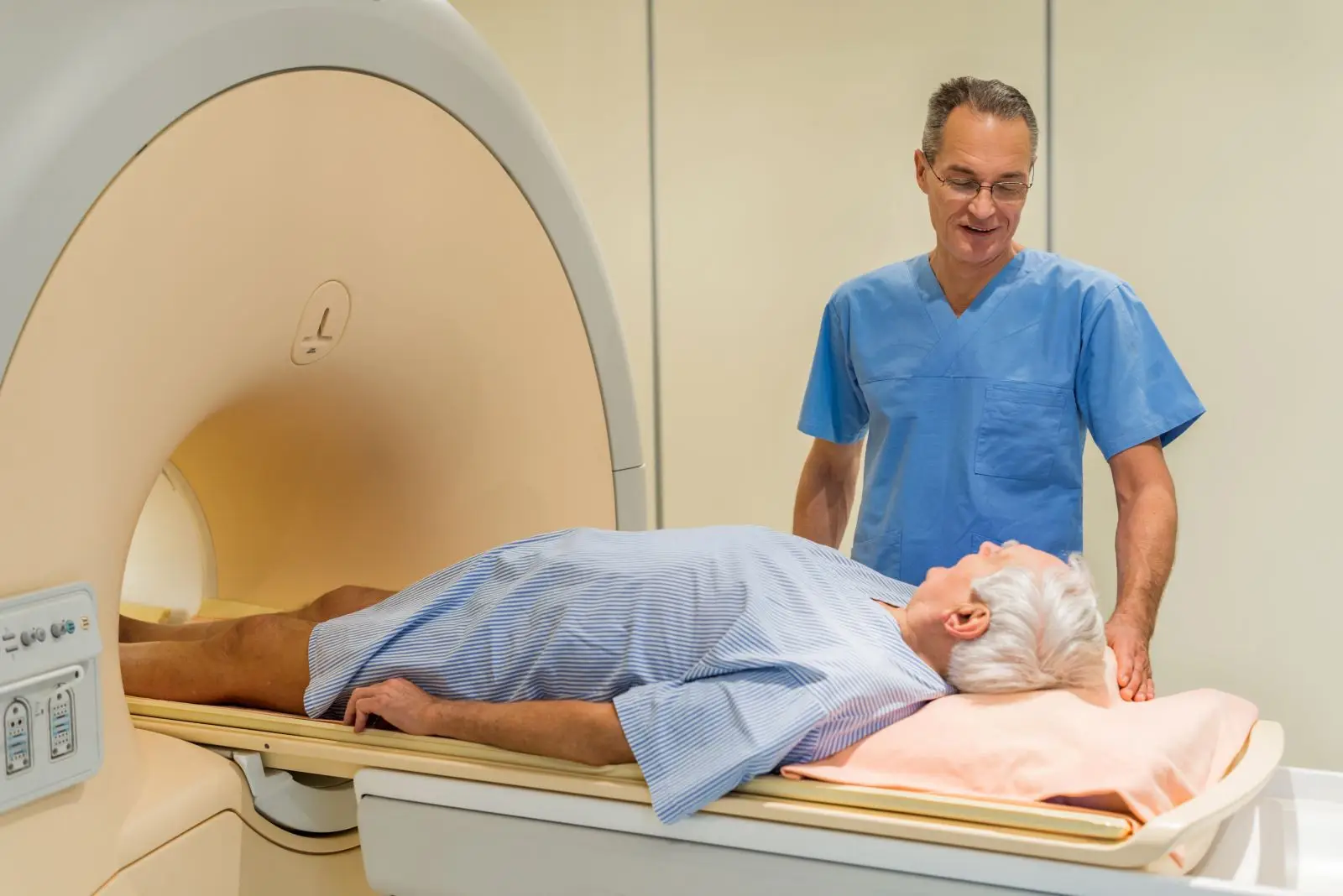
- MRI findings (including PI-RADS score and whether the lesion looks confined)
- Clinical stage (what the doctor feels on exam and what imaging suggests)
- Extent of cancer in the biopsy (positive cores, percent involvement)
- Your health and preferences (age, other conditions, goals, side effect tolerance)
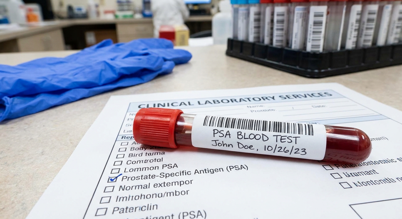
PSA adds context, but it is not a grade
PSA can rise for many reasons including benign prostate enlargement and inflammation. A higher PSA does not automatically mean higher-grade cancer, and a lower PSA does not guarantee low-grade cancer. What matters is how PSA fits with prostate size, MRI findings, and biopsy results.
What PI-RADS means
PI-RADS is a 1 to 5 scale radiologists use to describe how suspicious a finding is on prostate MRI. In general, higher numbers mean higher suspicion. PI-RADS does not diagnose cancer by itself, but it helps guide whether targeted biopsy is needed and how confident your team is that the biopsy sampled the most important area.
MRI can guide how confident we are in the biopsy
If an MRI showed a suspicious area and the biopsy sampled that lesion, doctors may feel more confident the biopsy captured the most important disease. If the MRI was concerning but the biopsy looks low-grade, your team may consider whether:
- the lesion was adequately targeted
- repeat biopsy is needed
- additional imaging or a second pathology review would help

Risk groups you may hear
Patients are often told their cancer is “low risk,” “intermediate risk,” or “high risk.” Intermediate risk is sometimes split into favorable and unfavorable. These groupings are based on a combination of Grade Group (or Gleason), PSA, and clinical stage, and sometimes MRI and biopsy volume. Grade Group is a major driver, but it is not the only driver.
Biopsy vs prostatectomy grades
One more nuance that can reduce confusion later: a biopsy samples parts of the prostate, not the whole gland. If someone has surgery, the final pathology from the prostatectomy specimen can sometimes show a different Grade Group than the biopsy did (either higher or lower). That does not mean the biopsy was “wrong.” It means we learned more once the entire prostate was examined.
Monitoring vs treatment
Every practice and every patient is a little different, but these are common patterns I see in shared decision-making.
When active surveillance is commonly considered
- Grade Group 1 (Gleason 3+3) with low volume disease
- Selected cases of Grade Group 2 (Gleason 3+4) when the pattern 4 component is small and other features are favorable
- PSA, PSA density, MRI, and biopsy volume that all point toward lower-risk cancer
Active surveillance is not “doing nothing.” It is a structured plan that typically includes PSA tests, periodic imaging, and repeat biopsy or repeat targeted sampling at intervals.
When active treatment is more often recommended
- Grade Group 3 (Gleason 4+3) or higher
- Higher PSA density or fast PSA rise that does not match the biopsy grade
- MRI features concerning for more extensive disease
- Higher volume cancer in cores, or additional adverse pathology features (for example IDC-P)
Treatment options may include surgery, radiation (with several approaches), and in some cases hormone therapy. The best choice depends on your risk category, anatomy, other medical conditions, and what side effects matter most to you.
Questions to ask
If you are holding a biopsy report right now, these questions can help you turn numbers into a plan:
- What is my Grade Group and what does it mean for my risk level?
- Is my Gleason score 3+4 or 4+3? How much pattern 4 is present?
- How many cores were positive, and what was the percent involvement?
- Did the report mention cribriform pattern or intraductal carcinoma (IDC-P)?
- What is my PSA density and how does it affect my options?
- How do my MRI findings, including my PI-RADS score, fit with the biopsy results?
- Am I a candidate for active surveillance? If so, what is the exact schedule?
- Should my biopsy slides be reviewed by a specialized genitourinary pathologist?
- If I choose surgery, how often do biopsy grades change on the final pathology?
When to call sooner
Most biopsy result discussions can safely wait for a scheduled follow-up, but contact your clinician promptly if:
- you have fever, chills, worsening pelvic pain, or feel acutely unwell after biopsy
- you cannot urinate
- you have heavy bleeding that is not easing
- you are feeling overwhelmed, panicked, or unable to function day to day while waiting for next steps
Your care team can address complications, speed up communication when appropriate, or connect you with support.
Bottom line
A Gleason score describes how aggressive prostate cancer looks under the microscope based on its most prominent patterns. Grade Groups translate that information into a simpler 1 through 5 scale. And while 3+4 and 4+3 both add up to 7, they do not carry the same risk because the dominant pattern is different.
If you feel stuck staring at the numbers, bring your report to your follow-up and ask your clinician to walk you through it line by line. You deserve to understand what you are being asked to live with, watch, or treat.