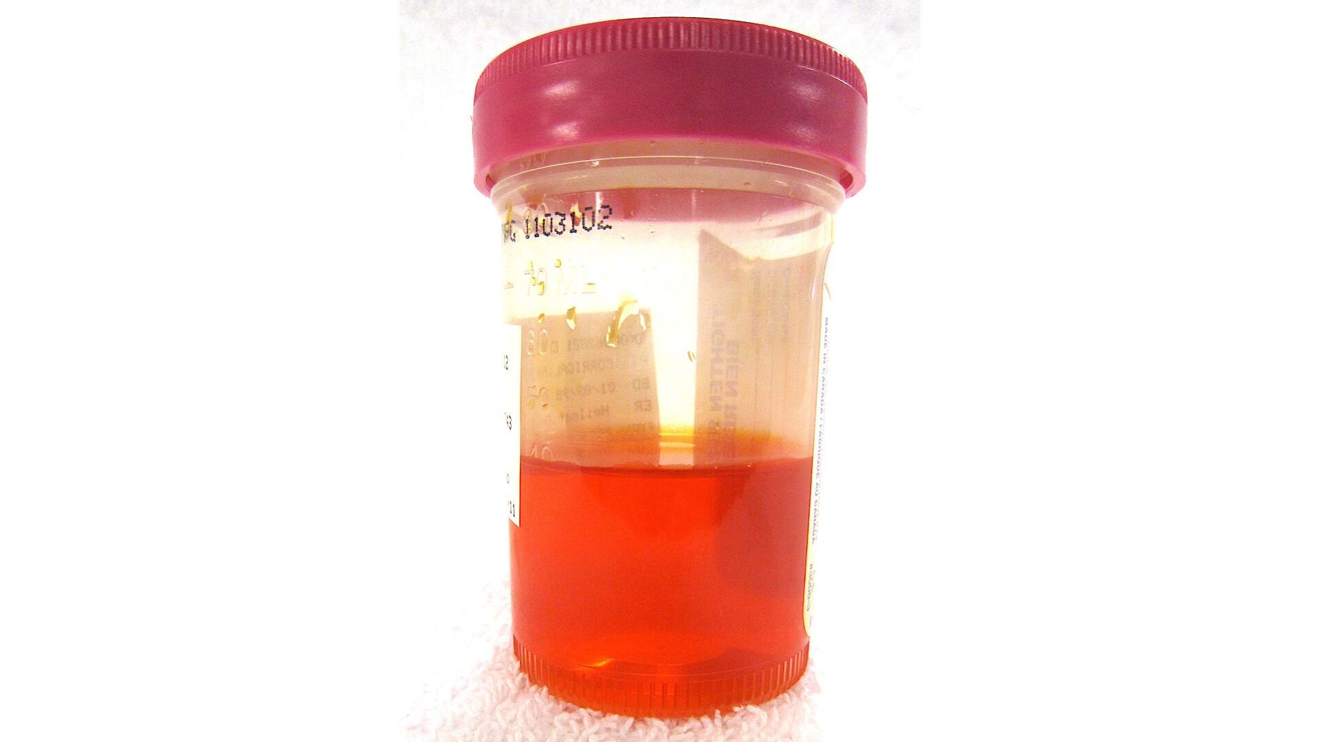
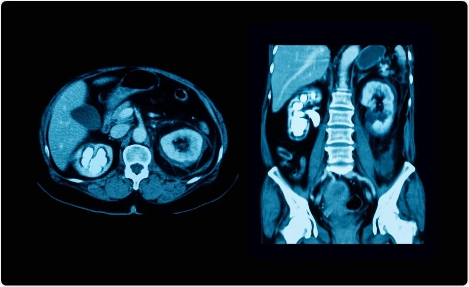
If you have blood in your urine or your clinician is concerned about a possible urinary tract tumor, you may be scheduled for a CT urogram. In plain terms, this is a specialized CT scan that takes timed pictures after IV contrast to look closely at the kidneys, ureters, and bladder and how well they become outlined on the delayed images.
I have found that people feel much calmer about this test once they understand two things: first, a CT urogram is not one single picture, it is a series of phases. Second, many results use very specific words like filling defect that sound scarier than they need to without context.
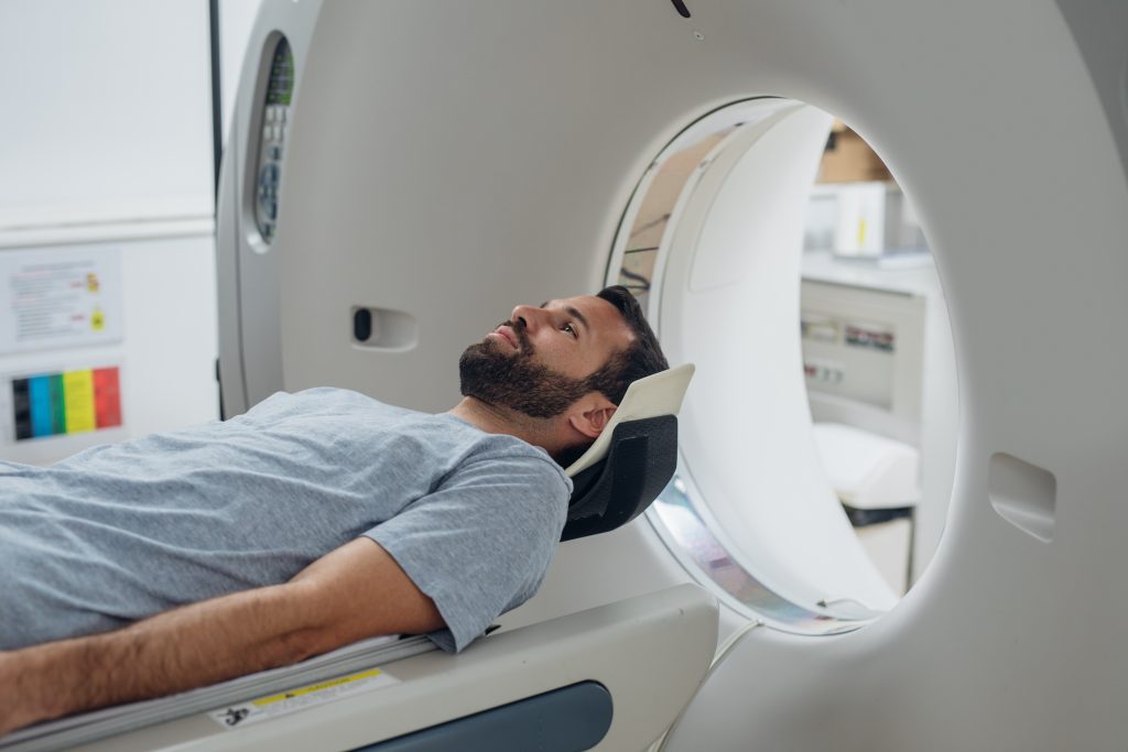
What a CT urogram looks for
A CT urogram is designed to evaluate the urinary tract in a way that standard CT scans do not always capture. It helps radiologists and urology teams look for problems that could explain bleeding or suspicious findings on other tests.
- Kidneys: the kidney tissue itself and the internal collecting system where urine begins
- Ureters: the thin tubes that carry urine from each kidney to the bladder
- Bladder: the lining and any areas that look irregular
It is often ordered when the goal is to rule out or evaluate issues involving the lining of the urinary tract, including growths, narrowing, or blockages.
CT urogram phases
Most CT urograms use iodinated IV contrast. The reason is straightforward: contrast moves through the bloodstream, into the kidneys, and then into the urine, which lets the scan highlight different structures at different times.
Protocols vary by facility and clinical question. Many centers use a combination of noncontrast, nephrographic, and excretory images, and some use a split-bolus approach to combine phases and reduce radiation dose.
1) Noncontrast phase
This is a scan done before the IV contrast goes in. It can help identify things that are naturally dense on CT, such as certain types of kidney stones or calcifications, and it provides a baseline to compare later images.
2) Corticomedullary phase
This is an early phase shortly after contrast injection. It highlights blood flow patterns in the kidneys. Not every facility includes this as a separate step for every patient, but when it is used, it can help characterize masses or vascular patterns.
3) Nephrographic phase
This phase is typically obtained when contrast has reached the kidney tissue more evenly. It is useful for evaluating the renal parenchyma, meaning the working tissue of the kidney where some masses are best seen.
4) Excretory phase
This is the hallmark of a CT urogram. After a delay, contrast has been filtered by the kidneys and is now in the urine. The collecting systems, ureters, and bladder can become outlined by contrast, helping radiologists look for:
- Areas that do not fill normally
- Any sudden narrowing or obstruction
- Abnormal lining irregularities
In some centers, you may be asked to change position, take a short walk, drink water, or have additional delayed images if the ureters have not filled well yet. Some protocols also use IV fluids or a small dose of a diuretic such as furosemide to improve ureteral filling, but this is not routine everywhere.
How to prepare
Your imaging center will give you instructions that match their protocol and your health history. These are the most common preparation steps patients ask me about.
Fasting and hydration
- You may be asked not to eat for a few hours beforehand, especially if IV contrast is planned.
- Hydration is often encouraged before and after unless your clinician has you on fluid restrictions. Being well hydrated can help you feel better after contrast and can improve urinary tract filling for images.
Full bladder
Some centers want you to arrive with a comfortably full bladder, while others prefer you empty right before scanning. Because this varies, I recommend calling ahead if you were not given specific instructions.
Medications
Most routine medications can be taken as usual, but you should confirm with your ordering clinician and the imaging center, especially if you take diabetes medications or have known kidney disease.
What to wear and bring
- Wear comfortable clothes that are easy to change out of if needed.
- Bring a list of allergies and your current medications.
- Be ready to answer questions about kidney function, prior contrast reactions, asthma, and thyroid disease.
What happens during the exam
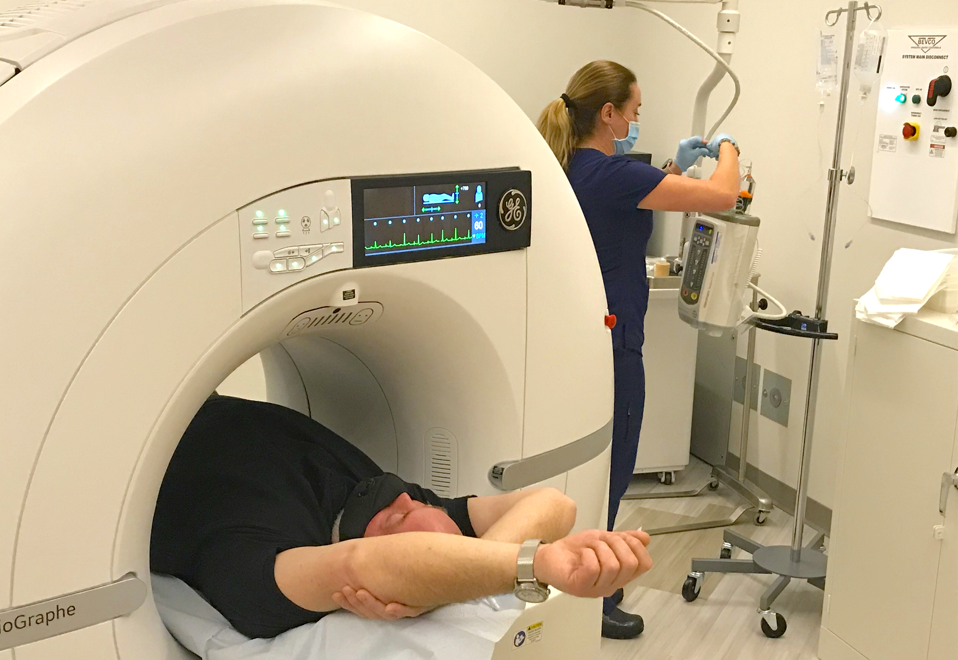
A CT urogram is usually an outpatient test. The scan itself is quick, but the visit is often longer because some images are taken after a waiting period for the excretory phase. In many departments, plan on roughly 30 to 90 minutes from start to finish, depending on how long it takes for the contrast to reach and outline the urinary tract.
- IV placement in your arm or hand.
- Initial scan may be done before contrast.
- Contrast injection. Many people notice a warm flush, a metallic taste, or a sensation of needing to urinate. These feelings are common and usually pass quickly.
- Additional scans are taken at planned time points.
- Delayed images may be done after waiting several minutes to allow contrast into the urine and improve ureter visualization.
It is important to hold still when asked and follow breathing instructions. Motion can blur the images, which sometimes leads to repeat scanning.
If you are anxious or claustrophobic, tell the technologist. CT scanners are relatively short and open compared with MRI, and most people are surprised by how fast it feels once it starts.
What “filling defect” means
When radiologists use the term filling defect, they are usually describing a spot where contrast filled urine should be, but something appears to be taking up space inside that contrast column.
Think of the excretory phase like dye outlining the inside of plumbing. If there is something inside the tube, such as a clot, a stone, a growth, or even an air bubble or artifact, the dye may flow around it and create a visible gap. That gap is the filling defect.
In the collecting system
A filling defect in the renal pelvis or calyces can be caused by several possibilities, including:
- Blood clot within the collecting system
- Stone or calcified material
- Tumor involving the lining of the collecting system (urothelial tumor)
- Sloughed papilla in certain kidney conditions
Radiologists look at the shape, margins, whether it moves between images, and whether it enhances like tissue to help narrow what it most likely represents.
In a ureter
The ureters are narrow, so even small issues can stand out. A ureteral filling defect may reflect:
- Stone (often associated with upstream swelling or delayed drainage)
- Blood clot
- Ureteral stricture or narrowing
- Urothelial tumor along the ureter lining
If the ureter did not fill well with contrast, a report may also mention nonopacification or limited evaluation. That does not automatically mean something is wrong. It may mean the timing, hydration status, or transient ureter spasm limited the view.
In the bladder
In the bladder, a filling defect can be related to:
- Blood clot sitting in the bladder
- Bladder stone
- Bladder mass or irregularity of the lining
Because clots can mimic masses, the next step is often based on how the finding looks and your overall clinical picture. Your clinician may recommend follow-up imaging or a direct look inside the bladder with cystoscopy.
Contrast safety
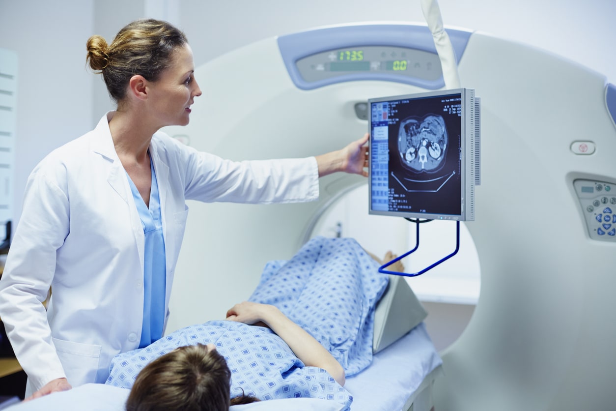
Most people receive iodinated IV contrast without any serious problem, but it is absolutely worth reviewing the main safety issues ahead of time. Your imaging team should screen you, but I always tell patients to speak up if any of the points below apply to them.
Kidney function
Iodinated contrast is cleared through the kidneys. If you have reduced kidney function, you may need recent blood work (often a creatinine and eGFR) before the exam. The main concern is the risk of kidney injury in people with significantly reduced eGFR and additional risk factors. For many patients with normal or mildly reduced kidney function, the risk is low, but your clinician should make that call for your situation.
Prior contrast reaction
If you have ever had hives, swelling, trouble breathing, or a significant reaction after CT contrast, tell the ordering clinician and the radiology center early. Depending on the severity, they may:
- Recommend a different imaging study
- Use a premedication plan
- Plan extra monitoring
A warm flush or metallic taste during injection is common and is not the same as an allergic reaction.
Asthma and severe allergies
People with asthma or a history of significant allergic reactions can have a higher risk of contrast reactions. This does not mean you cannot have contrast, but the team may take additional precautions.
Metformin
If you take metformin or certain combination diabetes medications, ask specifically what your facility recommends. Guidance often depends on your kidney function and whether you receive contrast. Do not stop medications on your own without instructions.
Thyroid disease
Iodinated contrast can affect the thyroid in certain situations. If you have hyperthyroidism, thyroid nodules being evaluated, or a history of thyroid storm, bring it up to your clinician.
Pregnancy and breastfeeding
CT uses ionizing radiation. If you might be pregnant, tell the imaging center before the scan. For breastfeeding, iodinated contrast is generally considered compatible in most cases. Some people choose to pump and discard milk for 24 hours for personal comfort, but it is usually not required. If this is a major concern for you, ask your clinician what they recommend for your situation.
Radiation questions
A CT urogram often involves more radiation than a single phase CT because it may include multiple passes. The benefit is the detailed view of the urinary tract that other tests may not provide. If you are concerned about radiation exposure, it is reasonable to ask:
- How many phases are planned for my scan
- If any phases can be omitted based on my history
- Whether a split-bolus protocol is an option
- Whether there is an alternative test that answers the same question
In radiology, the goal is to use the lowest dose needed to get a diagnostic answer.
Alternatives and next steps
A CT urogram is not the only way to evaluate blood in the urine. Depending on your age, risk factors, symptoms, kidney function, and pregnancy status, your clinician might consider:
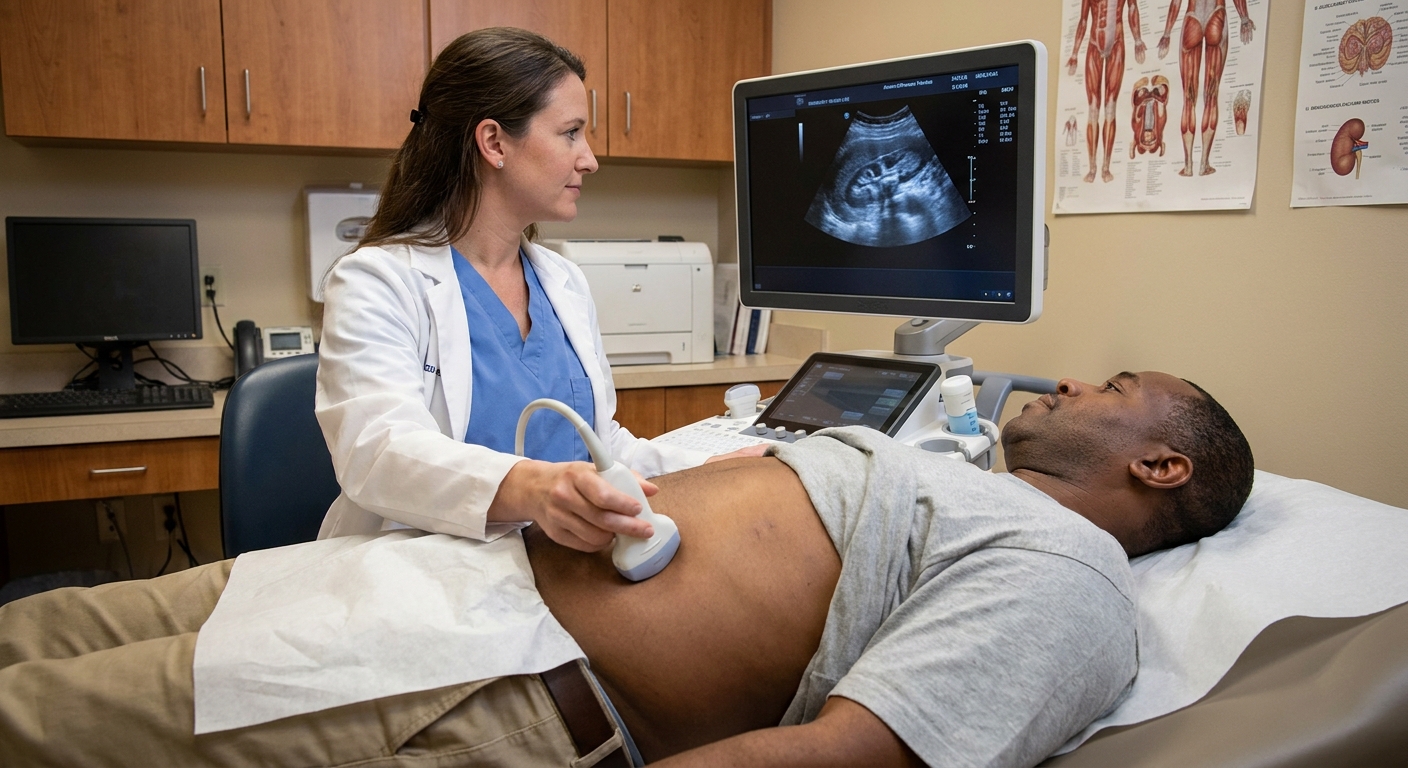
- Ultrasound: no radiation, useful for hydronephrosis and some kidney masses, but it may miss small urothelial lesions and small stones.
- MR urogram: uses MRI instead of radiation, can be helpful in selected patients, but availability and protocols vary.
- Cystoscopy: a urologist looks directly inside the bladder with a small scope. This is still commonly recommended for hematuria because CT can miss some small or flat bladder lesions.
If you are trying to make sense of why you need more than one test, this is often the reason: CT urogram and cystoscopy answer related but different questions.
After the test
Most people can return to normal activities right away.
- Drink fluids as directed, unless you have a fluid restriction.
- Watch for delayed reactions such as hives or itching later the same day. Seek urgent care for trouble breathing, facial swelling, or severe symptoms.
- Check your results plan. Ask who will call you, when you should expect results, and what the next step would be if something is seen.
Report terms
Reports can sound technical. Here are a few phrases you might see and what they generally refer to.
- Opacified: filled with contrast, meaning that segment was well seen.
- Nonopacified: not filled with contrast during the scan timing. This can be a limitation, or it can suggest an obstruction depending on the context.
- Hydronephrosis: swelling of the kidney collecting system, often from slow drainage or blockage.
- Urothelial thickening: the lining looks thickened. This can have multiple causes and is interpreted alongside symptoms and other findings.
- Filling defect: something appears to occupy space where contrast urine should be.
If you have your report in hand, I always recommend reading the Impression section first, then bringing questions back to your ordering clinician or urologist so the imaging findings can be matched to your specific situation.
When to get urgent help
Call your imaging center, clinician, or seek urgent care if you develop:
- Difficulty breathing, wheezing, or throat tightness
- Swelling of the lips, face, or tongue
- Widespread hives
- Severe dizziness or fainting
- Severe pain at the IV site with swelling or blistering
Quick FAQs
Is a CT urogram the same as a regular CT scan?
Not exactly. A CT urogram uses specific timing and delayed images after IV contrast to evaluate the urinary tract lining and how well the contrast outlines the collecting systems, ureters, and bladder.
Will I feel the contrast?
Many people feel a brief warm sensation, a metallic taste, or a feeling like they might urinate. These are common and usually pass within a minute or two.
If my report mentions a filling defect, does that mean cancer?
Not automatically. A filling defect means there is something within the contrast filled urine space that interrupts the outline. Causes can include clots or stones as well as tumors. The next steps depend on the size, shape, enhancement pattern, and your clinical history.
What if I have kidney disease?
Tell your clinician and the radiology team. You may need recent labs, and sometimes a different imaging approach is chosen to reduce risk.