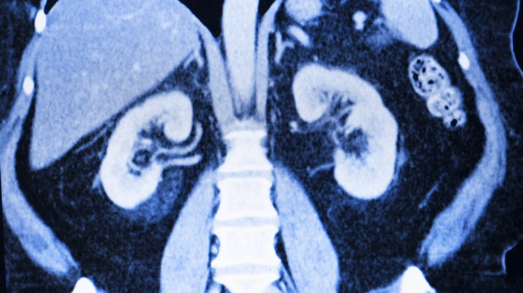
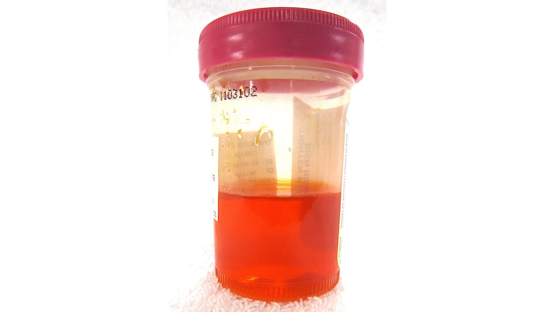
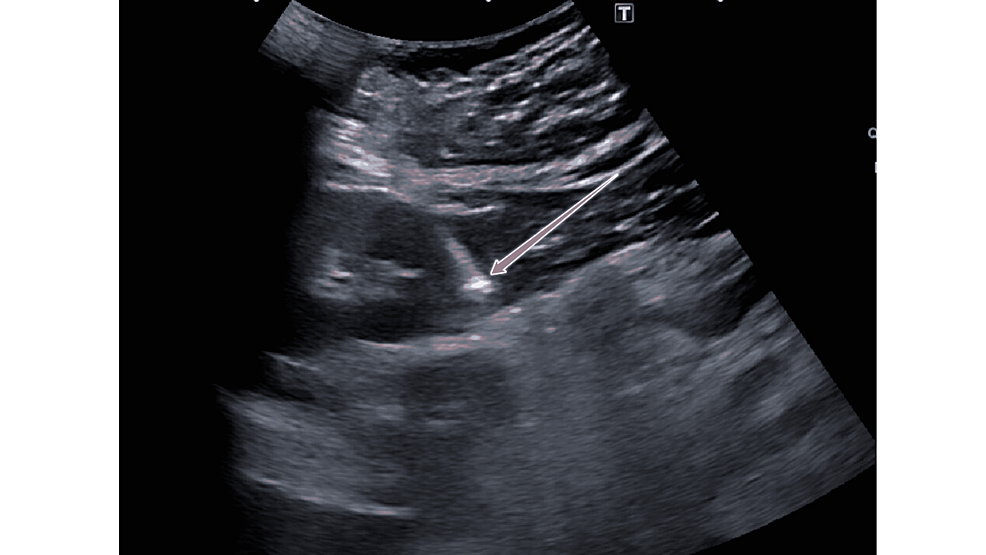
When a radiology report mentions an “angiomyolipoma,” many people understandably get stuck on one word: tumor. In family practice, I have walked patients through this exact moment, often after a scan done for flank pain, kidney stones, or blood in the urine. The reassuring news is that a renal angiomyolipoma, often shortened to AML, is usually benign. The important part is knowing which AML you have, how big it is, and whether it carries a meaningful risk of bleeding.

Renal angiomyolipoma, explained
A renal angiomyolipoma is a growth in the kidney made up of three tissue types:
- Angio = blood vessels
- Myo = smooth muscle
- Lipoma = fat
So an AML is essentially a mix of fat, muscle, and blood vessels in one lump. Most AMLs are found incidentally, meaning you were not looking for them and they showed up on imaging done for another reason.
Although AMLs are benign, they are not always “nothing.” The blood vessel component can be fragile, and in larger or higher-risk AMLs that can raise the risk of bleeding.
How imaging identifies AML
Many kidney masses look similar at first glance, which is why radiologists lean on certain identifying features. For AML, the key feature is often visible fat within the lesion.
CT scans
On CT imaging, macroscopic fat (fat you can clearly detect) inside a kidney mass strongly suggests an angiomyolipoma. When that classic fat finding is present, AML can often be diagnosed with high confidence without surgery.
A useful nuance is that radiologists do not rely on fat alone. They also consider the overall enhancement pattern and whether there are features that would be unusual for AML (for example, certain calcifications). Rarely, other kidney tumors can contain fat-like elements, and on occasion AML can coexist with another kidney lesion. This is why the final interpretation is always “in context.”
MRI
MRI can also detect fat within a lesion and help confirm the diagnosis, especially in people who cannot receive iodinated contrast for CT or when the CT findings are not straightforward. MRI may still use gadolinium contrast, and in people with advanced kidney disease that may be avoided or adjusted. Your imaging team will choose the safest option for you.
In clinic terms, if your report says something like “fat-containing renal mass consistent with angiomyolipoma,” that is typically the radiology team pointing out the hallmark that separates AML from many kidney cancers.

Symptoms
Small AMLs often cause no symptoms. When symptoms happen, they can overlap with other kidney problems, which is why AML is frequently discovered during a workup for:
- Blood in the urine, visible or only seen on a lab test
- Flank or back pain
- A feeling of fullness on one side (less common)
- Anemia if bleeding occurs
If a bleed is sudden and significant, people can develop severe flank pain, lightheadedness, weakness, or signs of shock. That is an emergency.
Why size matters
The main medical concern with AML is bleeding. Because AMLs contain abnormal blood vessels, some tumors can develop fragile areas that can leak or rupture.
Clinicians use a mix of factors to estimate bleeding risk, including:
- Tumor size
- Symptoms (pain, hematuria)
- Imaging features such as aneurysms within the tumor
- Growth over time
- Whether AMLs are multiple or associated with a genetic condition
You may hear a size threshold discussed in the range of about 4 cm as a point where bleeding risk becomes more clinically relevant for many patients, but it is not a universal rule. In practice, symptoms, tuberous sclerosis status, and the presence of an intratumoral aneurysm (often discussed when it is larger than about 5 mm) can matter as much as, or more than, diameter alone.
Possible AML bleed warning signs
Seek urgent care or emergency evaluation if you have any of the following, especially if you already know you have an AML:
- Sudden, severe flank or abdominal pain
- Fainting, dizziness, or severe weakness
- Fast heart rate, pale or clammy skin
- Visible blood in urine with worsening pain
AML and tuberous sclerosis
AMLs can occur as a single, sporadic finding, but they can also be associated with genetic syndromes, most notably tuberous sclerosis complex (TSC).
Sporadic AMLs are often solitary and discovered later in life, commonly in women. In TSC, angiomyolipomas are more likely to be:
- Multiple
- On both kidneys
- Detected at younger ages
- More prone to growth and complications
If you have multiple AMLs, have them at a young age, or have other features suggestive of TSC, your clinician may recommend a genetics evaluation or screening for other TSC-related findings.
Fat-poor AML
Not every angiomyolipoma has enough visible fat to “announce itself” on imaging. These are often called fat-poor AMLs or fat-invisible AMLs.
Here is the practical takeaway: when fat is not clearly seen, some AMLs can resemble renal cell carcinoma and other solid kidney tumors. In that situation, your care team may recommend additional evaluation such as:
- Repeat imaging with a specific CT or MRI protocol
- Follow-up scans over time to monitor growth
- A referral to urology
- Sometimes a kidney mass biopsy
That can feel frustrating, especially after hearing that AMLs are “benign.” But the caution is appropriate. The goal is to avoid missing a cancer that happens to look similar when the classic visible fat finding is absent.
AML vs Bosniak cysts
People often see multiple terms on imaging reports, and it is easy to blend them together. A helpful way to separate these two common categories is this:
- Angiomyolipoma is a solid mass with components like fat, muscle, and blood vessels.
- Kidney cysts are typically fluid-filled. Complex cysts may be graded using the Bosniak classification to estimate cancer risk.
A simple cyst is basically a small fluid pocket in the kidney and is extremely common. An AML is a different entity entirely. Sometimes imaging can show both in the same person, especially as we age, which is one reason radiology reports can feel so dense.
What happens next
Next steps depend on your symptoms, tumor size, and imaging certainty. In general, management may include:
Observation and follow-up imaging
Many small, asymptomatic AMLs are monitored periodically to ensure they remain stable. How often follow-up imaging is done varies by risk level and local practice. It is usually individualized based on size, growth, symptoms, and higher-risk imaging features such as aneurysms.
Urology referral
If the AML is large, growing, causing symptoms, associated with TSC, or uncertain on imaging, a urologist is typically involved.
Treatment options
When an AML is higher risk or actively causing problems, common options include:
- Selective arterial embolization to reduce blood flow to the tumor, often used to control bleeding or lower risk
- Nephron-sparing surgery (partial nephrectomy) in selected cases
- Thermal ablation in carefully selected situations
- mTOR inhibitors (for example, everolimus), particularly for some TSC-associated AMLs
Your clinician will discuss which approach fits your situation, and why.
AML and pregnancy
If you are pregnant or planning pregnancy, make sure your clinician knows you have an AML. Hormonal and blood vessel changes in pregnancy can increase bleeding risk in some cases, especially with larger or higher-risk lesions. This does not automatically mean something bad will happen, but it does mean your follow-up plan should be intentional.
Questions to ask
If you have been told you have a renal angiomyolipoma, these questions can make your next appointment much more productive:
- How big is it in centimeters, and where is it in the kidney?
- Does the imaging show visible fat, or is it considered fat-poor?
- Are there any aneurysms within it, and if so, how large are they?
- Do I need follow-up imaging? If yes, when and with what type of scan?
- What symptoms should prompt urgent evaluation?
- Do I have one AML or multiple? Does that raise concern for tuberous sclerosis?
- Do I need a urology referral?
When to seek care now
Please seek urgent evaluation if you have known AML and develop sudden severe flank or abdominal pain, fainting, significant weakness, or other signs that could suggest internal bleeding.
And if your first clue is blood in the urine, it is always worth getting checked even if it goes away. Many causes are benign, but hematuria can also be a sign of stones, infection, or a kidney mass that needs evaluation.
If you are holding a radiology report and feeling scared, take a breath. “Benign” does not mean “ignore,” and “tumor” does not automatically mean “cancer.” The safest path is understanding what type of lesion it is, confirming it confidently, and following it appropriately.
FAQ
Is a renal angiomyolipoma cancer?
Most renal angiomyolipomas are benign (non-cancerous). The main concern is bleeding risk in higher-risk tumors and making sure a look-alike cancer is not being missed in fat-poor cases.
Can an angiomyolipoma cause blood in the urine?
Yes. AMLs can sometimes cause hematuria (blood in the urine), especially if they bleed.
Do all AMLs need treatment?
No. Many are monitored. Treatment is considered when bleeding risk is higher, symptoms occur, growth is seen, or imaging cannot confidently rule out another diagnosis.
What does “fat-containing renal mass” mean?
It often means the imaging detected visible fat within a kidney lesion, which strongly suggests an angiomyolipoma in most cases. Your clinician will interpret this in context with size, symptoms, and other imaging features.