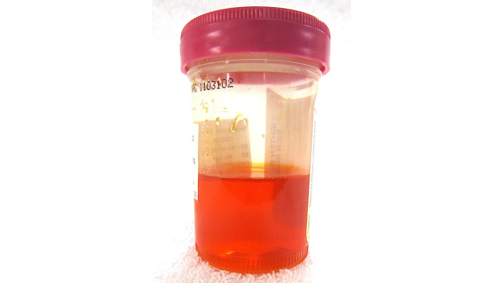
If you have blood in your urine, a nagging ache in your flank, or you were told a scan shows a “solid kidney mass,” it is completely normal for your mind to jump to the worst-case scenario. In clinic, one of the first things I do is slow the moment down and translate the terms so you can make decisions from a place of clarity, not panic.
Renal cell carcinoma, often shortened to RCC, is the most common type of kidney cancer in adults. Most RCCs arise from cells lining the kidney’s filtering tubes (the renal tubules), which are part of the nephron system that helps your body manage fluid, salts, and waste. Many kidney masses are found incidentally on imaging done for something else, and many are not cancer. But when RCC is on the list of possibilities, the next steps are about confirming what the mass is and choosing the right level of treatment for the stage and for your overall health.
What renal cell carcinoma is
Renal cell carcinoma is a cancer that begins in the kidney tissue itself, not in the urine-collecting space (the renal pelvis) and not in the bladder. The word “renal” means kidney. The word carcinoma means a cancer that arises from epithelial (lining) cells.
There are different subtypes of RCC, which matters because behavior and treatment can vary. The most common are:
- Clear cell RCC: the most common subtype.
- Papillary RCC: a less common subtype that can have different growth patterns.
- Chromophobe RCC: tends to be less common and often less aggressive.
Your care team may not know the subtype right away. Sometimes imaging suggests a pattern, but the subtype is confirmed when tissue is examined, either after surgery or after a biopsy when a biopsy makes sense for your situation.
Risk factors
Sometimes RCC shows up in people who did “everything right,” so I try to frame risk factors as context, not blame. Factors that can increase risk include:
- Smoking (current or past).
- Higher body weight and metabolic health factors.
- High blood pressure.
- Family history or inherited syndromes (for example, von Hippel-Lindau).
- Some workplace exposures (your clinician may ask about this depending on your history).
Common symptoms and why they happen
Early RCC often causes no symptoms. That is one reason kidney cancers are frequently found on scans done for other reasons.
Blood in the urine (hematuria)
Blood can be visible (pink, red, cola-colored) or only detected on a urine test. In RCC, bleeding can occur when a tumor irritates or invades small blood vessels that drain into the urine pathway.
Important: blood in the urine has many causes, including urinary tract infection, kidney stones, enlarged prostate, and other cancers such as bladder cancer. Any new blood in urine, especially if it is visible, should be evaluated promptly.
Flank pain
Flank pain is discomfort along your side or back, under the ribs. A kidney mass can cause pain by stretching the kidney’s capsule, pressing on nearby structures, or occasionally bleeding into the kidney area.
A mass seen on imaging
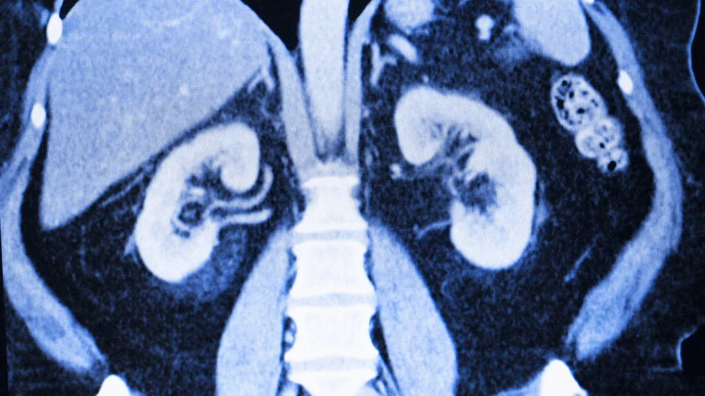
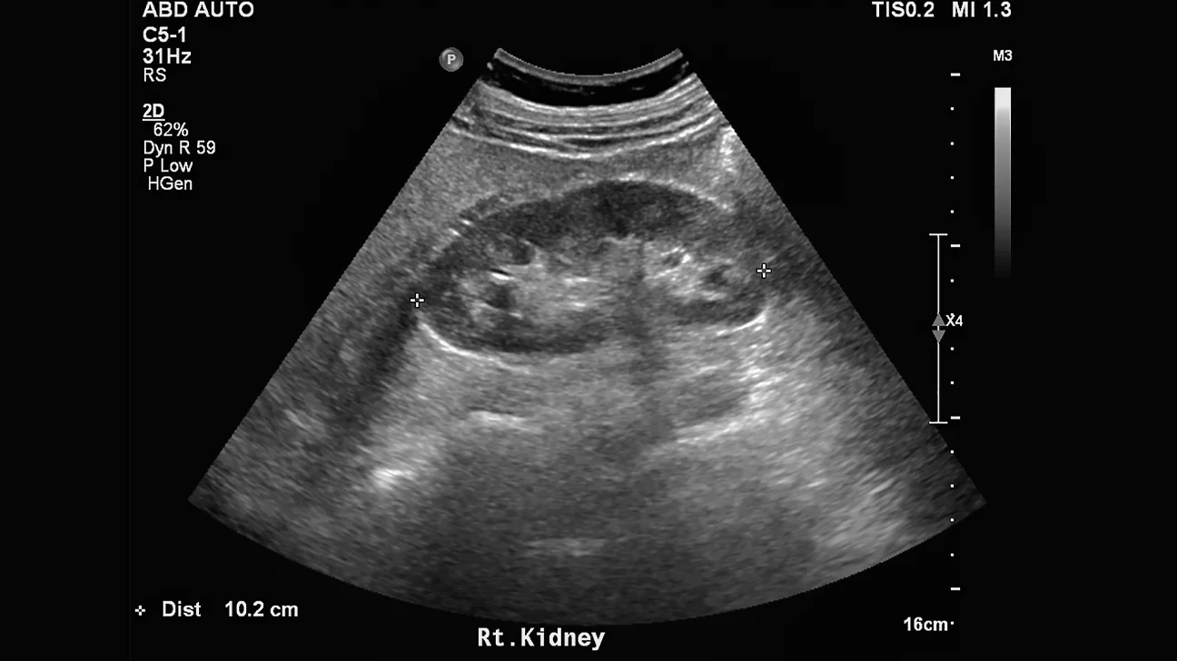
Many people first hear about RCC because an ultrasound, CT, or MRI report mentions a “solid enhancing renal mass.” The word enhancing means it takes up contrast dye on imaging, which often suggests blood supply within the lesion. Solid masses that enhance are more suspicious for RCC than simple fluid-filled cysts.
One nuance I often mention: not every enhancing mass is RCC. Some benign tumors (such as oncocytoma) can also enhance, which is one reason your clinician may talk about probabilities and next steps rather than making a diagnosis from imaging alone.
How doctors evaluate a suspected kidney cancer
The workup usually has two goals: (1) characterize what the mass is, and (2) determine whether it is confined to the kidney or has spread. Your exact pathway may differ based on what was seen on the first scan and whether you have symptoms.
Imaging tests
- CT scan with and without contrast (often a dedicated renal mass protocol): commonly used to assess size, enhancement, location, and whether nearby structures are involved.
- MRI: helpful when CT contrast is not safe for you, when the mass is complex, or when more detail is needed (for example, evaluating possible involvement of a vein).
- Ultrasound: often the first test that finds a mass, but CT or MRI is typically needed to better characterize it.
- Chest imaging (often CT or X-ray): may be used because the lungs are one place kidney cancer can spread.
Urine and blood tests
These do not diagnose RCC on their own, but they help assess kidney function, anemia, infection, and overall health status. Common examples include a urinalysis, complete blood count, and kidney function tests (creatinine and estimated GFR).
Is a biopsy always needed?
Not always. Some kidney masses go straight to treatment based on imaging and clinical context. A biopsy may be considered when:
- The imaging is not clearly cancer versus benign.
- You are considering active surveillance and want more information.
- You are considering ablation (freezing or heating the tumor), where tissue diagnosis can guide decisions.
- The mass might represent something other than RCC (for example, lymphoma or metastasis from another cancer), where treatment would be very different.
Biopsy has limitations. Sometimes a biopsy is nondiagnostic (not enough tissue, or tissue that cannot be interpreted). A small sample can occasionally miss cancer cells, and a “benign” result does not always fully settle the question if the sample was limited or the imaging looks highly suspicious. Your urologist and radiologist weigh these factors based on your case.
Understanding staging
Staging describes how large the tumor is and whether it has spread beyond the kidney. It is one of the biggest drivers of treatment choices.
Clinicians often describe stage in plain terms like this:
- Localized: confined to the kidney.
- Locally advanced: extends into nearby tissues or major veins, or involves nearby lymph nodes.
- Metastatic: has spread to distant organs such as lungs, bones, liver, brain, or distant lymph nodes.
Formally, staging is recorded using the TNM system (tumor size and extent, lymph nodes, and metastases). In many cases, the most definitive staging is finalized after surgery when pathology can examine the tumor directly, although imaging-based staging guides early decision-making.
Many RCCs are found when they are still localized, which opens the door to kidney-sparing approaches for the right person and the right tumor.
How stage guides treatment
When people hear “kidney cancer,” they often assume the only option is removing the whole kidney. In reality, there is a spectrum, and the best plan depends on tumor size, location, stage, symptoms, kidney function, and your overall health and preferences.
1) Active surveillance
Active surveillance means monitoring the mass with scheduled imaging and clinic visits rather than treating right away. It is most often considered when:
- The mass is small, often under about 4 cm.
- The tumor appears slow-growing.
- You have other medical conditions where surgery could carry higher risk.
- You have limited life expectancy or would not benefit from aggressive treatment.
This is not “doing nothing.” It is a structured plan. If the tumor grows more quickly than expected, changes in appearance, or causes symptoms, treatment can be revisited.
2) Ablation
Ablation uses needles placed into the tumor (usually guided by CT) to kill tumor cells with extreme cold (cryoablation) or heat (radiofrequency or microwave ablation). It is most commonly used for small, localized tumors and can be a good option when:
- You want to avoid or are not a good candidate for surgery.
- The tumor is small and located in a spot that can be safely targeted.
- Preserving kidney function is especially important.
Outcomes tend to be best for smaller tumors that are more peripheral or exophytic (growing outward). More central or hilar tumors can be harder to treat safely with ablation because of nearby blood vessels and the urine-collecting system.
Ablation can be very effective in carefully selected cases, but it may require close follow-up imaging and sometimes repeat treatment if any tumor persists.
3) Partial nephrectomy
Partial nephrectomy removes the tumor plus a margin of normal tissue while leaving the rest of the kidney in place. For many localized kidney cancers, especially smaller ones, this is preferred when technically feasible because it helps preserve kidney function.
Partial nephrectomy is often considered when:
- The tumor is localized and removable without sacrificing the whole kidney.
- You have only one kidney, reduced kidney function, diabetes, high blood pressure, or other risks for kidney disease.
- The tumor size and location make kidney-sparing surgery safe and reasonable.
4) Radical nephrectomy
Radical nephrectomy removes the entire kidney, and sometimes nearby tissue depending on the situation. It is more likely when:
- The tumor is larger or involves structures that make partial removal unsafe or incomplete.
- The tumor is located in a way that prevents a good margin while sparing the kidney.
- There is suspicion of more extensive local spread.
Many people live well with one kidney, but this option requires careful attention to your remaining kidney’s health over time.
Later-stage or metastatic RCC
If RCC has spread beyond the kidney, treatment often includes systemic therapy such as immunotherapy and targeted therapies. Today, combinations are common (for example, an immune checkpoint inhibitor paired with another immune drug or a VEGF-targeted therapy), and the plan is usually guided by a multidisciplinary team (urology, medical oncology, sometimes radiation oncology).
Radiation is not typically the main treatment for the kidney tumor itself, but it can be very useful for symptom control, such as treating painful bone metastases or certain brain metastases.

RCC vs kidney cyst grades
A common point of confusion is the difference between a solid kidney mass and a kidney cyst.
A simple kidney cyst is a fluid-filled sac and is extremely common, especially as we age. Simple cysts are usually benign and often need no treatment. Occasionally, a clinician will recommend follow-up if a cyst is not perfectly “simple” on imaging or if the report describes atypical features.
Some cysts are more complex and have features like septations (thin walls inside), thicker walls, or calcifications. Radiology reports may assign a risk category using a system called the Bosniak classification. In everyday terms:
- Lower-risk cyst categories typically mean the cyst looks mostly benign and may only need monitoring.
- Higher-risk cyst categories have features that make cancer more likely and may prompt surgical removal or closer evaluation.
RCC, by contrast, is most often a solid enhancing mass, not a simple fluid-filled cyst. A complex cyst can be cancer, but it is evaluated differently than a clearly solid mass.
RCC vs fat-containing benign masses
Another kidney finding that causes understandable anxiety is a mass that contains fat. The most classic benign fat-containing kidney tumor is an angiomyolipoma (AML).
Angiomyolipoma (AML) in brief
- AML is usually benign.
- It is made of a mix of blood vessels, smooth muscle, and fat.
- When it is small and not causing symptoms, it is often monitored.
Why it still matters is that AML can sometimes bleed, especially as it gets larger, and that bleeding can cause pain and blood in the urine.
Key difference: many AMLs have visible fat on imaging, which helps radiologists distinguish them from RCC.
One important nuance is that fat-poor AML exists. These AMLs do not show obvious fat on scans and can look more like RCC, which is why your report might recommend further imaging, biopsy, or a specialist visit even when a mass may ultimately be benign.
Questions to ask about a solid renal mass
When you are sitting with a report that feels like a foreign language, a few targeted questions can bring things into focus:
- How big is the mass (in centimeters), and where is it located in the kidney?
- Does it enhance with contrast? If yes, how strongly?
- Does it look more like RCC, AML, oncocytoma, or a complex cyst?
- Is there any sign it extends outside the kidney or involves lymph nodes or veins?
- What are my options: active surveillance, ablation, partial nephrectomy, radical nephrectomy?
- How will this choice affect my kidney function long-term?
- Do I need a biopsy? If not, why not?
- What follow-up schedule do you recommend for imaging and labs, and how might that schedule change after treatment?
When symptoms need urgent care
Most causes of blood in the urine are not immediately life-threatening, but some situations should be treated as urgent. Seek prompt care (urgent clinic, urgent care, or ER depending on severity) if you have:
- Blood in urine with clots or trouble urinating.
- Severe flank pain, especially with nausea or vomiting.
- Fever, chills, or feeling very ill along with urinary symptoms.
- Symptoms of significant blood loss such as lightheadedness, fainting, chest pain, or shortness of breath.
- Only one functioning kidney and new significant urinary symptoms.
Living in the in-between
Waiting for the next scan, the specialist appointment, or the biopsy result can be the hardest part. Two things can be true at once: this finding deserves a thorough evaluation, and many kidney masses are treatable, especially when found early.
At a high level, localized RCC is often curable with surgery or other localized treatments. More advanced disease is more complex, but it is increasingly treatable with modern systemic therapies. Your exact outlook depends on the stage, tumor biology, and your overall health.
If you have a copy of your report, it is reasonable to bring it to your appointment and ask your clinician to walk you through it line by line. You are not being difficult. You are being appropriately informed.
FAQ
Is renal cell carcinoma the same as bladder cancer?
No. RCC starts in the kidney tissue. Bladder cancer starts in the bladder lining and is evaluated and treated differently, even though both can cause blood in the urine.
Can a kidney stone cause the same symptoms as RCC?
Yes. Stones can cause blood in the urine and flank pain, often with sudden severe pain that comes in waves. Because symptoms overlap, imaging is important.
If the mass is small, does that mean it is not cancer?
Not necessarily. Small masses can be cancerous or benign. Size helps guide risk and treatment options, but it does not give a definitive answer by itself.
Will removing part of the kidney cure RCC?
For many localized RCCs, surgery (partial or radical nephrectomy) can be curative. The likelihood depends on stage, tumor features, and whether cancer is confined to the kidney.
If I have one kidney removed, what can I do to protect the remaining kidney?
Focus on the basics that truly move the needle: keep blood pressure controlled, manage diabetes if present, avoid smoking, stay well-hydrated unless your clinician advises otherwise, and review kidney-safe medication use (including anti-inflammatory drugs) with your healthcare team.