If you have ever read an imaging report that says something like “Bosniak IIF renal cyst” or “Bosniak III lesion,” you know how quickly a single number can raise your stress level. In clinic, I see this all the time: a patient feels fine, the scan was done for something else, and suddenly there is a classification that sounds ominous.
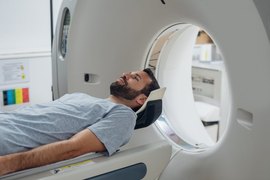
The good news is that the Bosniak classification is not a diagnosis of cancer. It is a standardized way radiologists describe kidney (renal) cystic masses seen on contrast-enhanced CT or MRI, based on how the cyst looks and whether it has features that can raise concern for malignancy. It helps your care team decide whether a cyst can be left alone, followed over time, or evaluated by a urologist for possible treatment.
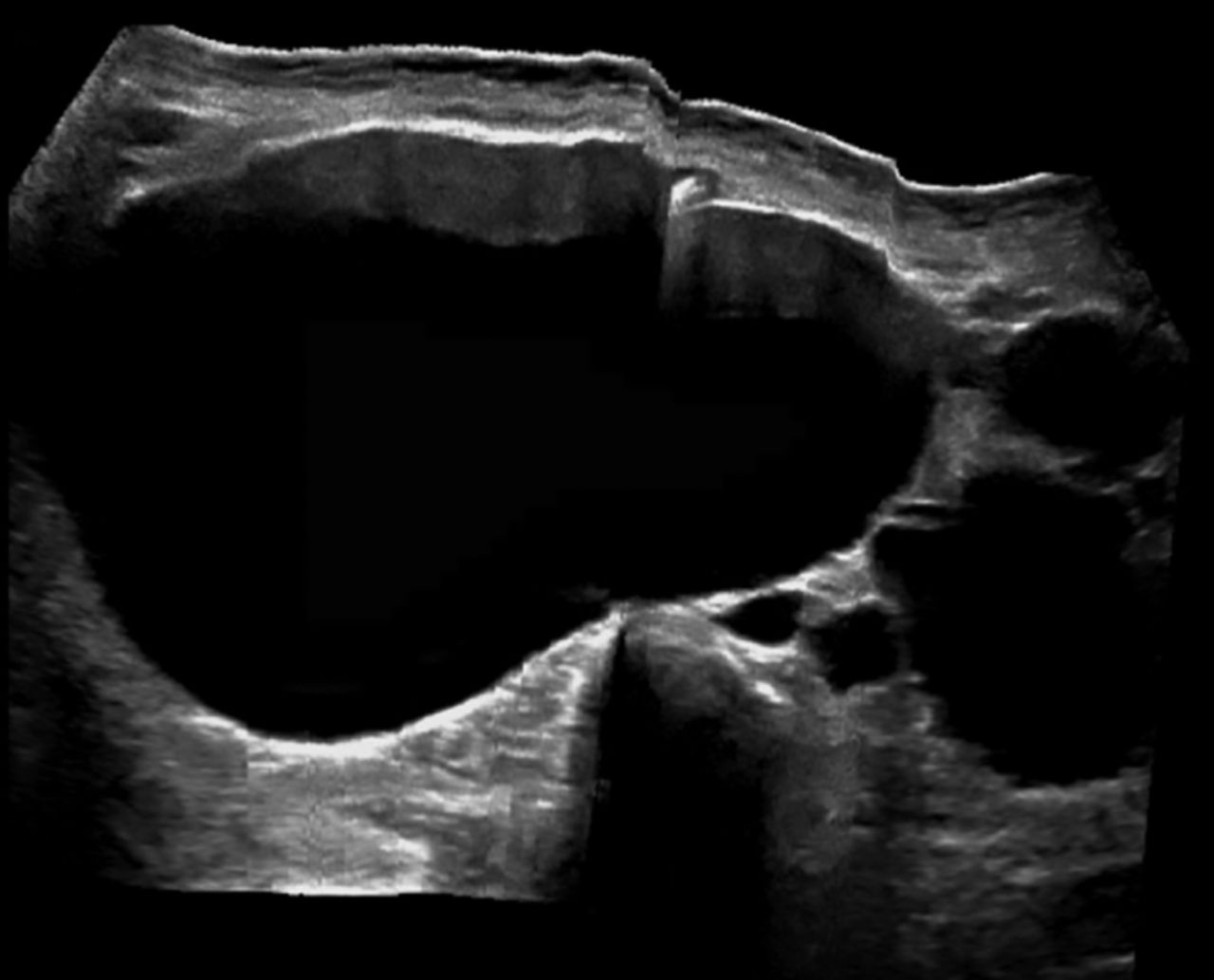
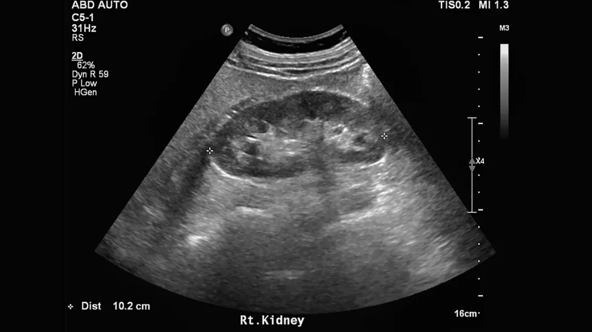
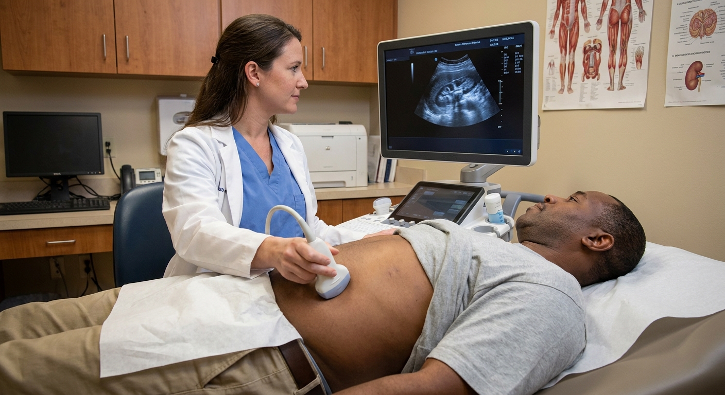
One quick clarification that helps: ultrasound can find kidney cysts, but Bosniak categories are generally assigned using CT or MRI with contrast, because contrast is what allows radiologists to assess enhancement.
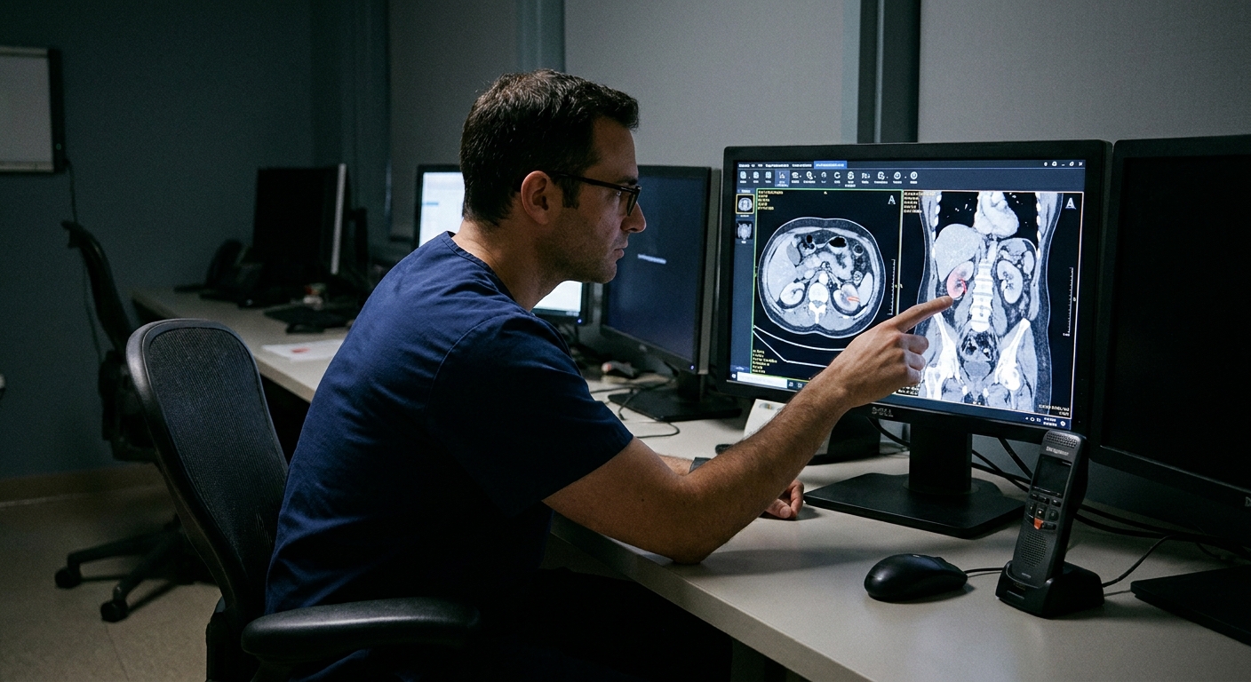
What it is and is not
The Bosniak classification sorts renal cystic masses into categories based on imaging features such as:
- Wall thickness and whether the wall looks smooth or irregular
- Septa (thin internal partitions) and how many there are
- Calcifications in the wall or septa
- Enhancement, meaning parts of the cyst take up contrast on CT or MRI (a key clue that vascular tissue may be present)
- Nodules or solid components inside the cyst
It is designed for cystic kidney findings. It is not used to grade hydronephrosis and it is not meant to sort out other look-alikes. It also does not apply to solid renal masses, which are evaluated differently.
It is also not a substitute for your clinician’s full picture of risk, which includes your age, symptoms, prior imaging, kidney function, family history, and more.
Why enhancement matters
A simple fluid-filled cyst can look bright or dark depending on the modality, but the key point is this: a simple cyst does not enhance after contrast. When the radiologist sees enhancing tissue in a cyst wall, septum, or nodule, that suggests vascular tissue rather than just fluid. That finding can increase concern for a malignant lesion.
Radiologists are careful about this. On CT, enhancement is often assessed by comparing attenuation (Hounsfield units) before and after contrast. On MRI, radiologists may use subtraction images and other sequences to confirm true enhancement and avoid false alarms.
This is one reason your report might recommend MRI for further characterization if a CT finding is borderline.
If you cannot receive contrast (for example, because of a prior severe contrast reaction or certain kidney function concerns), your team may use alternative strategies. Just know that non-contrast scans can be limited for Bosniak classification because enhancement is such a central feature.
Bosniak categories
Below is a practical, patient-centered way to understand each category. Exact percentages vary across studies, by whether the older system or Bosniak v2019 criteria were used, and by which lesions went to surgery. Think of these as general risk trends, not promises.
Bosniak I: simple cyst
What it looks like: A thin, smooth wall. No septa. No calcifications. No enhancement.
What it implies: Benign. This is the classic common kidney cyst many adults develop over time.
Typical plan: No follow-up imaging needed unless you have symptoms or your clinician has another reason for monitoring.
Approximate cancer risk: Essentially 0%.
Bosniak II: minimally complex
What it looks like: May have a few thin septa or fine calcifications. No suspicious enhancement or solid nodules.
What it implies: Very low risk of cancer.
Typical plan: Usually no follow-up needed.
Approximate cancer risk: Essentially 0%.
Bosniak IIF: follow-up
The “F” is for follow-up.
What it looks like: More complexity than category II, such as multiple thin septa, thicker walls or septa, more extensive calcification, or subtle features that are not clearly worrisome but are not confidently benign either. Enhancement, if present, is not in a clearly high-risk pattern.
What it implies: Low but not negligible risk. Many IIF cysts never change. The purpose of follow-up is to confirm stability over time.
Typical plan: Surveillance imaging at set intervals. Protocols vary, but a common approach is repeat imaging at about 6 months and 12 months, then annually for up to about 5 years if stable, depending on local practice and your overall situation.
What “good news” looks like: No growth, no new enhancement, no new nodules. Stability over time strongly supports benign behavior.
Approximate cancer risk: Often cited around 5% to 10%, with variability across studies.
Bosniak III: indeterminate
What it looks like: Thicker or irregular walls or septa that enhance after contrast. It often looks more complex or irregular than a benign cyst, but imaging alone cannot always provide a final answer.
What it implies: Meaningful risk of malignancy. Some Bosniak III lesions are benign, but many are not.
Typical plan: Referral to a urologist. Management is individualized. Surgery (often partial nephrectomy when appropriate) is still common, but active surveillance is increasingly used in selected patients, especially for smaller lesions, slower-growing lesions, or when other health factors make intervention higher risk.
Approximate cancer risk: Often cited around 40% to 60%, depending on criteria and selection.
Bosniak IV: clearly suspicious
What it looks like: A cystic mass with enhancing soft tissue nodules or solid components, in addition to complex walls or septa.
What it implies: Highest likelihood of malignancy among Bosniak categories.
Typical plan: Urology referral for definitive management. Treatment is individualized, but surgery is commonly recommended when feasible. Size, growth rate, kidney function, and your overall health all factor into the plan.
Approximate cancer risk: Often cited around 80% to 90% or higher.

Watched vs treated
As a general rule:
- Watched: Bosniak IIF (and sometimes category II if there is an unusual clinical reason)
- Usually referred to urology for possible treatment: Bosniak III and IV
- Typically no action needed: Bosniak I and II
There are exceptions. For example, an older adult with significant medical problems may choose surveillance even for a higher-category lesion if the risks of surgery outweigh benefits. On the other end, a younger person with a growing Bosniak IIF cyst that begins to show more convincing enhancement or a new nodule may be managed more aggressively.
Why you may see v2019
You may see references to an updated system, often called Bosniak v2019. Radiology groups refined the criteria to improve consistency across readers and imaging methods, especially with MRI.
For patients, the most important takeaway is simple: the category is still meant to communicate how concerning the imaging features are and what the next step should be.
Common questions
Does a higher category mean cancer?
No. It means the imaging features look more suspicious and the chance of cancer is higher. Bosniak III is “indeterminate” for a reason, and final diagnosis often requires pathology if surgery is performed.
If I feel fine, can I ignore it?
Most kidney cysts cause no symptoms, even when they need monitoring. Decisions here are based on imaging features and risk, not just how you feel. If your cyst is Bosniak IIF, III, or IV, do not ignore it. Make sure there is a clear follow-up plan.
Why MRI after CT?
MRI can sometimes better characterize enhancement and internal complexity without additional radiation. It can help confirm whether a cyst truly fits a higher-risk category or whether it is less concerning than it first appeared.
Can these cysts go away?
Some cysts remain stable for years. True regression can happen but is less common. The more important goal is verifying stability and catching meaningful change early.
Next steps checklist
If you have a Bosniak category on your report, these steps tend to reduce confusion and prevent missed follow-up:
- Ask what imaging was used (CT vs MRI) and whether contrast was given.
- Confirm the exact category (II vs IIF matters).
- Ask what features drove the category (enhancement, thick septa, nodules).
- Ask whether the scan was non-contrast. If it was, ask if that limits classification and whether a contrast CT or MRI is recommended.
- Get a clear follow-up timeline in writing if you are in Bosniak IIF.
- Ask if urology referral is recommended now (typically for Bosniak III or IV).
- Ask how size affects the plan, especially for Bosniak III or IV, since size and growth rate often influence whether surveillance, ablation, or surgery makes the most sense.
- Bring prior scans if you had imaging at another facility. Stability across years can be very reassuring.
If you are ever unsure what the plan is, I encourage you to ask one direct question: “What is the next step, and when should it happen?” That single sentence prevents many delays in care.
When to seek urgent care
A Bosniak category itself is not usually an emergency. But seek urgent evaluation if you have:
- Severe flank or abdominal pain that is new or worsening
- Fever or chills with urinary symptoms
- Visible blood in the urine
- Inability to urinate, or symptoms of severe dehydration
Those symptoms can point to issues like infection, stones, or bleeding that deserve prompt care regardless of cyst category.