When someone comes into clinic worried about blood in the urine, one of the most common fears I hear is: “Does this mean cancer?” Often, the answer is no. Still, visible blood in the urine tends to carry a higher cancer risk than microscopic blood, especially in older patients and people with higher-risk histories (like smoking). So when your clinician is checking for urothelial cancer (cancer that starts in the lining of the urinary tract), they may order a test called urine cytology.
Urine cytology is not the same as a urine culture (which looks for bacteria) or a routine urinalysis (which checks for blood, protein, and other markers). Instead, it is a lab test where a specialist looks at cells that have shed into your urine and checks whether they look suspicious for cancer, especially high-grade urothelial cancer.
What urine cytology looks for
The inside lining of your bladder, ureters, and the kidney’s collecting system (the renal pelvis and calyces) is made of urothelial cells. These cells can shed naturally into urine. If a tumor is present, abnormal cells may also shed and be carried out in the urine stream.
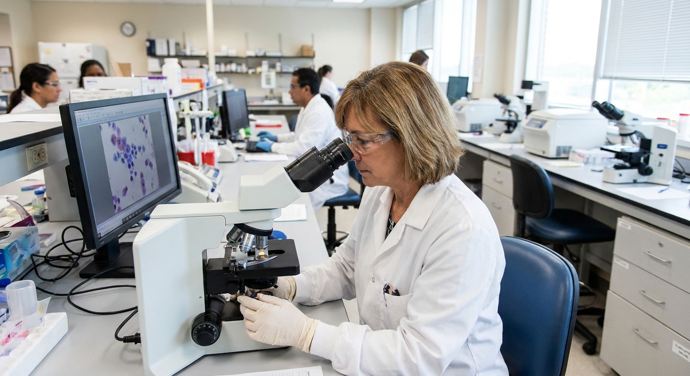
In urine cytology, a cytopathologist examines these cells under a microscope, focusing on whether they look like high-grade (more aggressive) cancer. In plain language, they are looking for cells that look clearly “off,” such as cells with very enlarged, irregular-looking nuclei and other features that suggest high-grade disease.
What it does not do
- It does not rule out cancer by itself. A normal result cannot fully exclude urothelial cancer, especially low-grade tumors.
- It does not pinpoint a location. Abnormal cells may come from the bladder or elsewhere in the urinary tract.
- It is not a biopsy. It looks at shed cells, not a piece of tissue.
How the sample is collected
There are two common ways urine is collected for cytology. The method matters because it affects how many cells are available for the lab to evaluate.
Voided urine sample
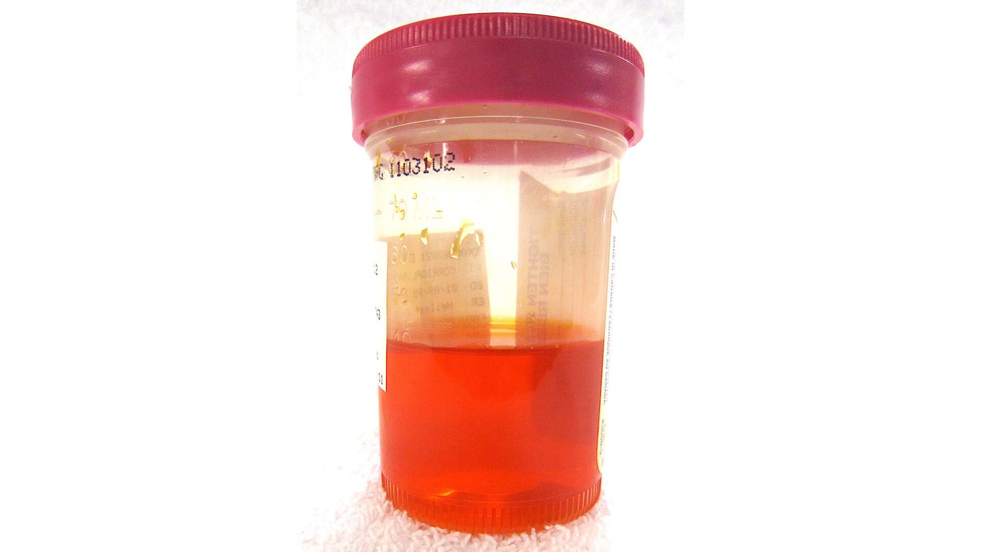
A voided sample is simply urine you pass into a sterile cup. Your clinician may ask for:
- A single sample, or
- Samples on multiple days, since cancer cells may not shed consistently
Practical tip: Many labs prefer a freshly collected sample brought in promptly. If you are given instructions about timing and delivery, follow them closely so cells do not degrade before they are processed.
Washings during a scope
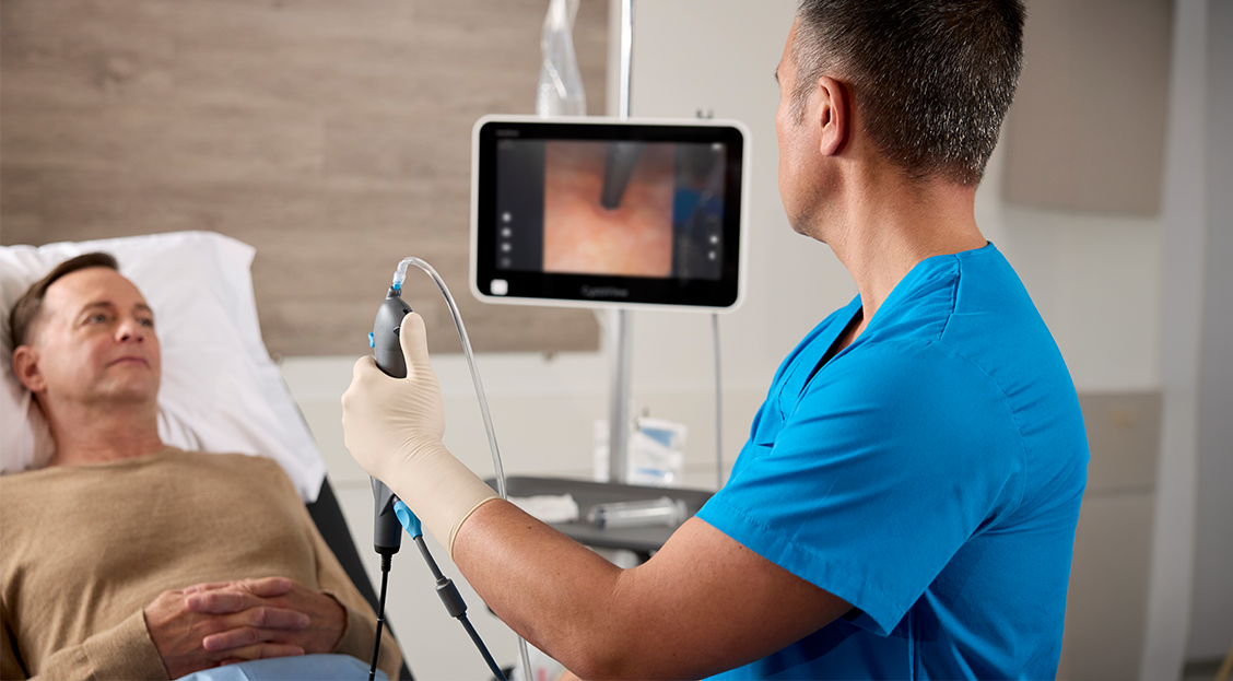
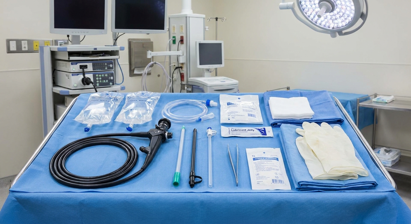
Sometimes cytology is collected as a “washing” during a scope procedure. The most common example is a bladder washing during cystoscopy, where sterile fluid is introduced into the bladder and then collected back for analysis. In selected cases, clinicians may also collect upper-tract washings during procedures like ureteroscopy (from the ureter or renal pelvis).
Compared with a voided specimen, washings can provide a higher-cellularity sample, which may improve the chance of detecting abnormal cells. The yield still varies based on technique, lab processing, and tumor characteristics.
Understanding the results
Lab wording can vary, but urine cytology results generally fall into a few broad categories. Many labs follow the Paris System, so you may see terms like “Negative for high-grade urothelial carcinoma,” “Atypical urothelial cells,” “Suspicious for high-grade urothelial carcinoma,” or “High-grade urothelial carcinoma.” Your urologist or primary care clinician should interpret the result in the context of your symptoms, risk factors, and other testing.
Negative
A negative result means the lab did not see malignant-appearing cells in the sample provided. On some reports, this may be phrased as “negative for high-grade urothelial carcinoma.”
Important: A negative cytology is better at lowering concern for high-grade cancer than it is at ruling out all bladder tumors. Many low-grade tumors shed cells that look fairly normal, so cytology can miss them.
Atypical or suspicious
These results mean some cells looked abnormal, but not abnormal enough for the lab to confidently call cancer. This can happen for several reasons, including:
- Inflammation or infection
- Recent urinary tract instrumentation (for example, a catheter)
- Kidney stones or significant irritation
- Low cell count in the sample
“Atypical” does not automatically mean cancer, but it often leads to closer follow-up and, in many cases, repeat testing or additional evaluation.
Positive
A positive result suggests the lab saw cells that look consistent with high-grade urothelial cancer or another malignancy.
This can be emotionally jarring to read. What I tell patients is this: cytology is a strong warning sign, but it is not the final diagnosis. False positives are uncommon, but they can happen, particularly in the setting of severe inflammation, stones, or recent instrumentation. The next step is typically to find the source and confirm it with tissue.
Why positives need confirmation
Urine cytology examines cells floating in urine. It cannot show the architecture of a tumor, how deep it grows, or exactly where it is coming from. Because treatment decisions depend on these details, clinicians usually need tissue confirmation, often through biopsies taken during urologic evaluation.
In other words, cytology can raise or lower suspicion, but a diagnosis and staging typically require direct visualization and sampling of the urinary tract lining.
If your cytology comes back positive, ask your clinician: “What is the plan to confirm where these cells are coming from?” That question keeps the focus on the next clear step.
Why negatives can miss tumors
One of the most confusing parts of this test is hearing, “Your cytology was negative,” and then learning you still need additional evaluation. This is because urine cytology is much more sensitive for high-grade disease than for low-grade disease.
Low-grade urothelial tumors often shed cells that do not look dramatically abnormal under a microscope. In addition, tumors may not shed cells steadily, so the sample collected on one day may simply not contain representative cells.
This is exactly why clinicians use urine cytology as one piece of a bigger evaluation, not the whole answer.
How it fits into the workup
Urine cytology is commonly ordered when clinicians are evaluating:
- Visible blood in the urine (red or cola-colored urine)
- Microscopic blood found on testing
- Persistent urinary symptoms in higher-risk patients, such as long-term smokers or those with certain chemical exposures
- Surveillance after a history of bladder cancer, depending on the risk category and care plan
In modern practice, next steps are often guided by risk stratification based on factors like age, smoking history, and whether hematuria is visible or microscopic. That is why two people with the same cytology result may be advised to do different follow-up testing.
Keeping the tests straight
People often mix up which test does what. Here is a simple way to separate them:
- Urine cytology: looks at cells shed into urine.
- Cystoscopy: a urology procedure that lets the clinician look inside the bladder.
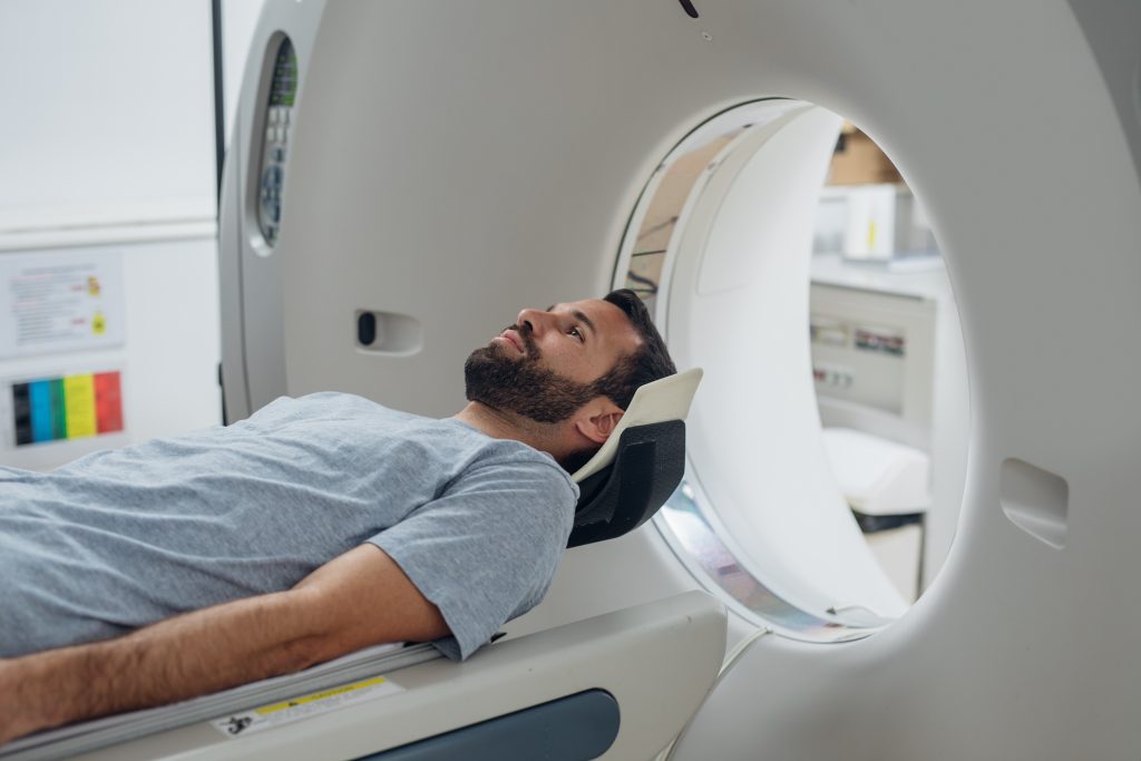
- CT urogram: a specialized imaging study that evaluates the kidneys and urinary tract using contrast imaging.
Each test answers a different question. It is very common for a clinician to recommend more than one, especially if there is blood in the urine or other red flags.
Common follow-up steps
Exactly what comes next depends on your situation and what has already been done, but common next steps after an abnormal cytology include repeat cytology, cystoscopy (if not already performed), imaging of the upper urinary tract, and sometimes ureteroscopy to directly evaluate the ureter and renal pelvis.
How to prepare
Preparation is usually minimal, but follow the instructions from your clinic or lab. Some general expectations:
- You may be asked to provide a urine sample at a lab or clinic.
- You might be asked for more than one sample on different days.
- If you recently had a urinary procedure or active infection, your clinician may time the test to reduce “atypical” results from irritation.
How long results take
Turnaround time depends on the lab, but results often take a few business days. Cytology requires specialized review, so it may not be as fast as a basic urinalysis.
Questions to ask
- Was my sample a voided specimen or a washing?
- Did the report say negative, atypical, suspicious, or positive (or Paris System wording like “negative for high-grade”)?
- Given my history and symptoms, how much does this result change my risk?
- What is the next step to confirm findings if the result is abnormal?
- If the result is negative, what tests are still needed and why?
When to seek urgent care
Even though urine cytology is often part of a planned evaluation, some symptoms should not wait:
- Inability to urinate
- Heavy bleeding or blood clots in urine
- Severe flank or abdominal pain with fever
- Fever, chills, and burning urination that suggest a spreading infection
If you are unsure whether your symptoms are urgent, call your clinician, an on-call nurse line, or seek prompt in-person care.
Bottom line
A urine cytology test is a microscope-based lab test that looks for abnormal urothelial cells in your urine. It is most helpful for detecting high-grade urothelial cancer, especially when there is visible blood in the urine or other risk factors. A positive cytology result typically needs tissue confirmation because cytology is not a biopsy. And a negative result does not fully rule out bladder tumors, particularly low-grade ones, which cytology can miss.
If you are in the middle of a hematuria workup, you are not alone. Ask for clear next steps and a timeline. Understanding what each test can and cannot tell you is one of the best ways to protect your peace of mind while still taking your health seriously.