Hearing the words “bladder cancer” can feel like the floor drops out from under you. In my clinic, one of the first things I do is slow things down and translate the medical language into something you can actually use. Many bladder cancers are diagnosed at an earlier stage, and there are effective treatments, but the best plan depends a lot on the stage and grade. Understanding the “what” and “what happens next” can help you walk into appointments with steadier footing.
What bladder cancer is
Your bladder is a hollow organ in your pelvis that stores urine. Bladder cancer starts when cells in the lining of the bladder begin to grow out of control. Most bladder cancers begin in the innermost lining that is constantly exposed to urine.
Bladder cancer is not one single disease. It includes different cell types and a range of how deeply the cancer has grown into the bladder wall. Those two details drive most treatment decisions.
Main types of bladder cancer
The “type” refers to what the cancer cells look like under a microscope.
- Urothelial carcinoma (transitional cell carcinoma): The most common type in the United States and many other regions. It starts in the urothelial cells that line the inside of the bladder.
- Squamous cell carcinoma: Less common in many countries, and often linked to long-term irritation or inflammation of the bladder. In some parts of the world, infection with schistosomiasis is a major risk factor for this type.
- Adenocarcinoma: Rare. It starts in gland-like cells.
- Small cell (neuroendocrine) carcinoma and other rare types: Uncommon, often more aggressive, and typically treated with specialized approaches.
If you only remember one thing: most people are dealing with urothelial carcinoma, and many of those cases are found before the cancer has invaded the bladder muscle.
Non-muscle-invasive vs muscle-invasive
The bladder wall has layers. A concrete way to picture it is: the inner lining, a supportive layer underneath it (often called the lamina propria), and then the bladder muscle.
Non-muscle-invasive bladder cancer (NMIBC)
Non-muscle-invasive means the cancer is in the inner lining and may have grown into the layer just beneath it, but it has not grown into the bladder muscle. This category includes:
- Ta: on the surface lining
- T1: into the connective tissue under the lining, but not muscle
- CIS (carcinoma in situ): a flat, high-grade cancer on the lining
NMIBC is often treated with procedures done through the urethra (no external incision) and, in many cases, medication placed directly into the bladder. It can come back, so follow-up is very structured.
Muscle-invasive bladder cancer (MIBC)
Muscle-invasive means the cancer has grown into the muscle layer of the bladder wall (stage T2 and above). Some cancers grow beyond the bladder into nearby structures (later T stages), and some have already spread to lymph nodes or other organs (metastatic disease). Those situations are treated differently, which is why staging details matter.
It is completely reasonable to ask your clinician: “Is this non-muscle-invasive or muscle-invasive?” That one sentence clarifies the roadmap.
Stage vs grade
These two words get used a lot, and they mean different things:
- Stage describes how deep the tumor has grown and whether it has spread.
- Grade describes how abnormal the cells look under the microscope and how aggressively they tend to behave (often simplified as low-grade vs high-grade).
Common symptoms
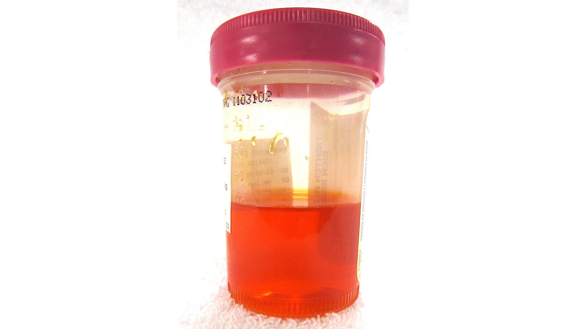
The symptom that brings many people in is blood in the urine, which can look pink, red, cola-colored, or sometimes be invisible and only show up on a urine test.
Other possible symptoms can include:
- Burning or pain with urination
- Needing to urinate more often
- Sudden urgency to urinate
- Feeling like you cannot empty your bladder fully
These symptoms can also be caused by infections, stones, enlarged prostate, or other non-cancer conditions. Still, visible blood in the urine should always be evaluated, especially in adults, because it is one of the clearest warning signs we do not want to ignore.
How bladder cancer is found
Diagnosis often happens in steps. Not everyone needs every test, but these are common parts of a bladder cancer workup:
- Urinalysis and urine culture: checks for blood and infection.
- Urine cytology: a lab looks for cancer cells shed into the urine. This can be more helpful for higher-grade cancers, but it can miss low-grade tumors.
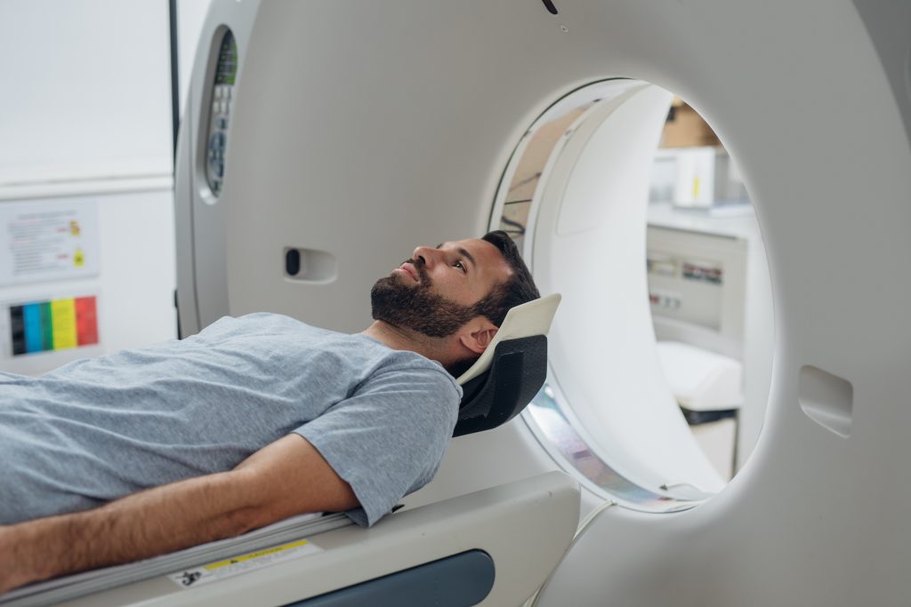
- Imaging: many people have a CT urogram or other imaging to evaluate the urinary tract, including the kidneys and ureters (the tubes that drain urine from the kidneys to the bladder).
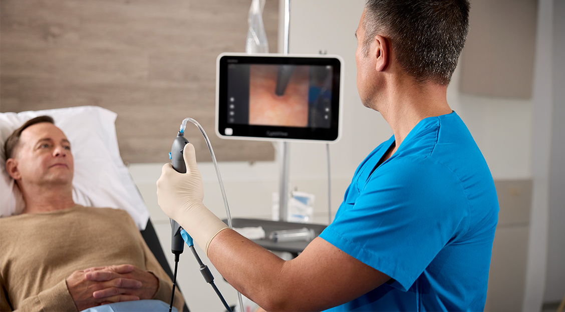
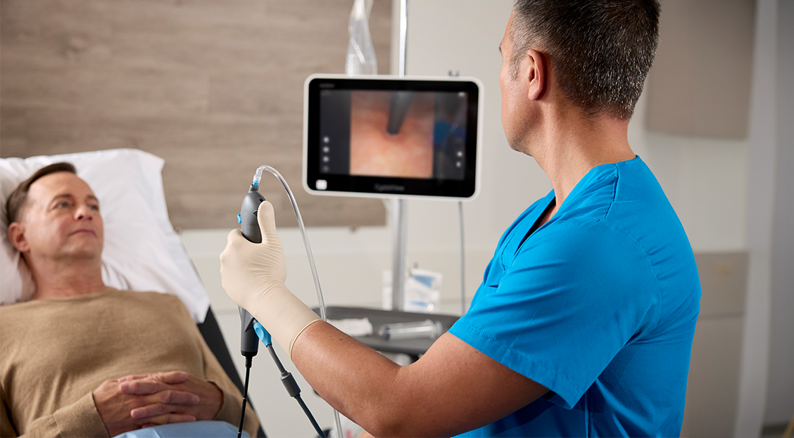
- Cystoscopy: a urologist looks inside the bladder with a small camera. This is the key test for directly seeing a bladder tumor.
Cystoscopy
Cystoscopy is an office procedure (sometimes done in a procedure suite) where a thin scope is passed through the urethra to look at the bladder lining.
What many patients want to know is: will it hurt? People describe it as pressure, a strong urge to urinate, or brief burning. A numbing gel is typically used, and the scope is often flexible for comfort. The actual viewing portion is usually quick.
Helpful tips I share with patients:
- Ask whether you should take acetaminophen or ibuprofen ahead of time, if you can safely take them.
- Plan for mild burning with urination and a small amount of blood afterward for a day or so.
- Call your clinician if you develop fever, worsening pain, or you cannot urinate after the procedure.
TURBT
If cystoscopy shows a suspicious growth, the next step is often a procedure called TURBT, which stands for transurethral resection of bladder tumor. Many people call it “scraping,” but it is more accurate to say the surgeon removes the tumor through the urethra using specialized instruments.
TURBT serves two purposes:
- Treatment: removes visible tumor tissue.
- Diagnosis and staging: the tissue is sent to pathology to determine the type, grade (how aggressive it looks), and how deep it has grown.
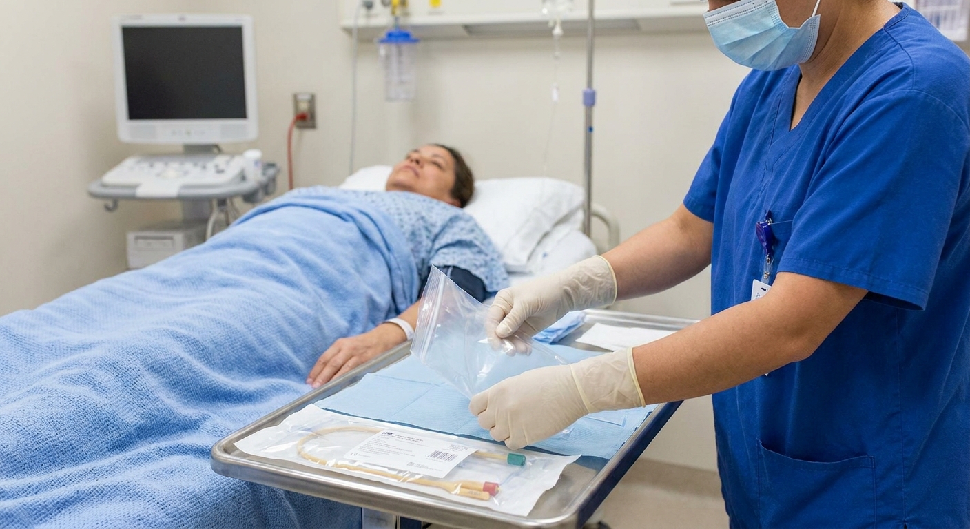
TURBT is commonly done with anesthesia. Some patients go home the same day. A temporary catheter is sometimes used afterward.
One important detail: for some people, a second TURBT is recommended. This is especially common for high-grade T1 tumors, and when the first specimen did not include bladder muscle, because we want to be confident about staging and that the tumor was fully removed.
What happens next
In many clinics, pathology results are reviewed within about 1 to 2 weeks, and your urology team uses that report to map out the next steps. Depending on your risk category, that may mean a repeat TURBT, an intravesical treatment plan, and a schedule for follow-up cystoscopies. Timelines vary by office, so it is fair to ask, “When should I expect results, and who will call me?”
Intravesical therapy
Intravesical therapy means medication placed directly into the bladder through a catheter. This is different from IV chemotherapy. The goal is to treat the bladder lining while limiting whole-body side effects.
Two common intravesical approaches are:
- Intravesical chemotherapy: In select low or intermediate-risk cases, a single dose may be placed into the bladder soon after TURBT (often within 24 hours) when it is safe to do so. It is typically avoided if there is concern for bladder perforation or significant bleeding.
- BCG (Bacillus Calmette-Guérin): A standard treatment for high-risk non-muscle-invasive cancers, including CIS. It helps your immune system attack cancer cells in the lining. It is commonly given as an induction course and, when tolerated and available, maintenance treatments over time.
During instillation, the medication is placed into the bladder and held for a prescribed time (often about 1 to 2 hours) before you urinate it out.
Common side effects include urinary burning, urgency, and frequency for a day or two. With BCG, flu-like symptoms can occur. Your urology team will give you specific safety instructions about urinating afterward and when to call for help.
Treatment paths
Treatment is personalized, but most care falls into a few broad paths based on stage, grade, and your overall health.
If it is non-muscle-invasive
- TURBT to remove the tumor and determine stage and grade
- Risk-based intravesical therapy (none, chemotherapy, or BCG depending on recurrence risk and aggressiveness)
- Surveillance with repeat cystoscopies because recurrence is common even when treatment is successful
If it is muscle-invasive
- Systemic therapy such as chemotherapy and/or immunotherapy, often before surgery
- Radical cystectomy (removal of the bladder) with urinary diversion in many cases
- Bladder-preserving approaches in select patients, often combining maximal TURBT with radiation and chemotherapy
If it is advanced or metastatic
If cancer has spread beyond the bladder and nearby lymph nodes, treatment usually centers on systemic therapy (immunotherapy, targeted therapy when appropriate, and sometimes chemotherapy), clinical trials when available, and symptom-focused care as needed. If you are in this category, you still deserve clear options and a plan that matches your goals.
If you are feeling overwhelmed, focus on the next decision point rather than the entire journey at once. Ask your clinician to write down your stage and grade and the recommended next step.
Risk factors
Bladder cancer often develops from a mix of exposures, age-related changes, and chance. Having a risk factor does not mean you caused your cancer, and many people diagnosed have no obvious risk factor.
- Smoking: The biggest known risk factor. Tobacco chemicals concentrate in urine and irritate the bladder lining over time. If you smoke, quitting after diagnosis can still matter. It can lower the risk of recurrence and supports overall health during treatment.
- Certain workplace chemical exposures: Historically linked to industries involving aromatic amines (for example, parts of dye, rubber, leather, and chemical manufacturing).
- Older age and male sex: Risk increases with age and is more common in men, though it occurs in all genders.
- Chronic bladder irritation: In some settings, long-term catheter use, recurrent bladder stones, or ongoing inflammation may increase risk.
- Prior pelvic radiation or certain chemotherapy drugs: Risk can be higher after pelvic radiation, and after medicines such as cyclophosphamide.
- Family history and genetic factors: Usually a smaller piece, but important to mention to your clinician.
Questions to ask
- What type of bladder cancer do I have (urothelial, squamous, other)?
- Is it non-muscle-invasive or muscle-invasive?
- What is the stage?
- What is the grade (low-grade or high-grade)?
- Did the pathology include bladder muscle in the specimen, and was it involved?
- When should I expect pathology results, and who will contact me?
- What is the plan for follow-up cystoscopies, and how often?
- Do you recommend intravesical therapy such as chemotherapy or BCG?
- Should I see a medical oncologist or radiation oncologist as part of my care team?
When to seek urgent care
Call your clinician or seek urgent care promptly if you have:
- Heavy bleeding with clots or inability to urinate
- Fever, chills, or feeling acutely ill after a urinary procedure
- Severe pain that is new or worsening
If you are newly diagnosed, please hear this clearly: you do not have to memorize everything at once. Bring a support person if you can, ask for your results in writing, and take it one step at a time. That is not just emotionally easier, it is often the best way to make solid medical decisions.