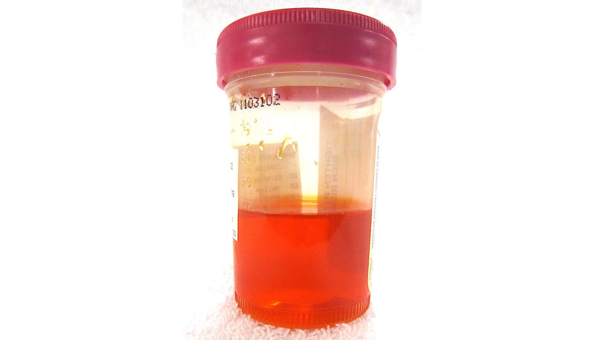
In clinic, one of the most important moments is when we realize we are not dealing with “blood in the urine” as a single symptom, but a whole pattern that points to a specific part of the urinary system. Nephritic syndrome is that kind of pattern. It is a set of findings that strongly suggests inflammation inside the kidney’s filters, not a problem in the bladder, ureters, or a simple kidney stone.
If your urine has turned tea or cola-colored, your blood pressure is creeping up, and lab work shows your kidney function is worsening, clinicians start thinking less about where urine exits the body and more about where it is made and filtered: the glomeruli.

Nephritic syndrome, defined
Nephritic syndrome is a clinical syndrome caused by inflammation of the glomeruli, the tiny filtering units inside your kidneys. When these filters are inflamed, they become “leaky” in a particular way, letting red blood cells slip into the urine and disrupting the kidney’s ability to regulate fluid balance, blood pressure, and waste removal.
It is called a syndrome because it describes a pattern that can be triggered by multiple different diseases. Two well-known examples are IgA nephropathy and lupus nephritis, but there are other causes as well.
What the classic pattern looks like
Nephritic syndrome is often recognized by a combination of symptoms and lab findings. Not everyone has every feature, and some people have only microscopic hematuria (blood seen on testing, not by eye). The overall cluster is what matters.
Dark, smoky urine
This color change is often due to blood coming from the kidney filters. When red blood cells are exposed to the urine’s chemistry and time in the urinary tract, the color can darken, creating a smoky, brown, tea, or cola appearance. Some people have obvious color change (gross hematuria), while others do not.
Rising blood pressure
Inflamed glomeruli can reduce effective filtration and activate hormonal systems that retain salt and water. That extra volume can push blood pressure higher, sometimes quickly.
Falling kidney function
Lab tests may show a rise in serum creatinine and a drop in estimated glomerular filtration rate (eGFR). Some people feel fine initially, which is why lab work is so important when urine changes appear.
Some protein in the urine
Protein leakage is common, but in nephritic syndrome it is typically less dramatic than in nephrotic syndrome. You might see protein on a dipstick or an elevated protein-to-creatinine ratio, typically not in the nephrotic range. Swelling can happen, but it is often milder than the heavy, generalized edema people associate with nephrotic syndrome.
Red blood cell casts on urinalysis
This is a strong clue that the problem is in the kidney itself. Red blood cell casts form when red blood cells and protein become molded together inside the kidney’s tubules. When we see casts, we think, “this bleeding happened inside the kidney,” not in the bladder.
One important nuance: casts are not always present, and they can be missed depending on timing and how the sample is handled. Another classic clue of a glomerular source is dysmorphic red blood cells (misshapen RBCs) on urine microscopy.

Why a stone or bladder workup can miss it
Blood in the urine can come from anywhere along the urinary tract, including:
- Kidneys (filters and tissue)
- Ureters (tubes from kidneys to bladder)
- Bladder
- Urethra
A kidney stone or bladder issue can certainly cause hematuria, but nephritic syndrome is different because it reflects filter inflammation.
Here is the practical takeaway: if a person has dark, tea-colored urine plus high blood pressure, reduced kidney function, and a urinalysis that shows protein and glomerular clues (like casts or dysmorphic RBCs), it is not enough to stop after evaluating for a stone or a bladder source. You can have a normal bladder ultrasound and still have significant kidney filter disease.
Common causes
Nephritic syndrome can be triggered by different conditions that inflame the glomeruli. Clinicians usually categorize causes by timing, associated symptoms, and blood and urine test patterns.
Examples (not a complete list)
- IgA nephropathy: often associated with episodes of blood in the urine, sometimes after an upper respiratory infection.
- Lupus nephritis: kidney inflammation related to systemic lupus erythematosus (SLE), often accompanied by other autoimmune symptoms and characteristic blood tests.
- Infection-related glomerulonephritis: can occur with or after infections, classically following strep throat or skin infections, and in adults sometimes during an active infection.
- ANCA-associated vasculitis: inflammation of small blood vessels that can involve the kidneys and other organs.
- Anti-GBM disease: may be kidney-limited or involve both kidneys and lungs. When lungs are involved, people may cough up blood.
The exact cause matters because treatment varies widely, from careful monitoring and blood pressure control to immune-suppressing medications.
How doctors evaluate it
If nephritic syndrome is on the table, the goal is twofold: confirm that the kidney filters are involved and identify the underlying cause quickly.
Urine testing
- Urinalysis with microscopy to look for red blood cells, protein, casts, and dysmorphic RBCs
- Urine protein-to-creatinine ratio (or albumin-to-creatinine ratio) to quantify protein loss
Blood testing
- Creatinine and eGFR to assess kidney function
- Electrolytes, including potassium and bicarbonate
- Complement levels (C3, C4) in some cases
- Autoimmune and inflammation markers when indicated (for example ANA, anti-dsDNA, ANCA)
- Infection-related tests when recent infection history suggests it
Blood pressure and fluid status
Blood pressure trends, weight changes, and swelling (especially around the eyes, hands, and ankles) help gauge fluid retention.
Imaging
Ultrasound is often used to evaluate kidney size and rule out obstruction. Imaging is helpful, but it does not replace urine microscopy when glomerular disease is suspected.
Kidney biopsy (sometimes)
A kidney biopsy is not always required, but it is often the most direct way to identify the exact type of glomerulonephritis and guide treatment, especially if kidney function is worsening, the diagnosis is unclear, or the presentation suggests a rapidly progressive process.

Treatment basics
Treatment depends on the cause and severity, but early management often focuses on protecting the kidneys while the diagnostic workup is underway.
- Blood pressure control: often with ACE inhibitors or ARBs when appropriate, because they can reduce pressure inside the glomeruli and lower protein loss. In acute kidney injury, pregnancy, significant hyperkalemia, or a rapidly rising creatinine, they may be deferred or used cautiously with close lab monitoring.
- Managing fluid overload: diuretics may be used if there is swelling or shortness of breath from fluid retention.
- Salt reduction: lowering sodium can help control swelling and blood pressure.
- Avoiding kidney stressors: nonsteroidal anti-inflammatory drugs (NSAIDs) and dehydration can worsen kidney function in vulnerable patients.
- Cause-specific therapy: some forms require immune suppression (like corticosteroids or other agents), while others improve with supportive care and time.
If you are being evaluated for nephritic syndrome, ask your clinician what your creatinine and eGFR trend looks like and whether your urine microscopy showed casts or dysmorphic red blood cells. Those details often clarify the level of urgency.
When to seek urgent care
Blood in the urine is never something to ignore, but certain signs raise the urgency because they suggest kidney involvement or rapidly changing kidney function.
Seek urgent or emergency care if you have:
- Dark, tea or cola-colored urine plus new swelling (face, eyelids, legs) or shortness of breath
- Severe headache, chest pain, confusion, or very high blood pressure readings
- Decreased urination or inability to keep fluids down
- Blood in the urine with fever and feeling very unwell
- Known kidney disease with a sudden change in urine color, blood pressure, or labs
If symptoms are milder but persistent, make an appointment promptly. A simple urine dip alone is not the full story. The combination of urine microscopy and kidney function labs is what helps separate a lower-tract issue from nephritic syndrome. If kidney function is worsening quickly, clinicians often involve nephrology early because some forms can progress rapidly and need urgent evaluation.
Nephritic vs. nephrotic
These two are easy to mix up because both involve the kidney filters.
- Nephritic syndrome: inflammation, blood in the urine (sometimes tea-colored), red blood cell casts or dysmorphic RBCs, rising blood pressure, and reduced kidney function.
- Nephrotic syndrome: heavy protein loss, significant swelling, low blood albumin, and often high cholesterol. Blood in the urine may or may not be present.
Some kidney diseases can have overlapping features, which is why your clinician focuses on your specific lab pattern rather than a name alone.
Questions I hear often
Can nephritic syndrome go away on its own?
Some causes improve with supportive care and time, especially if the trigger was an infection and kidney function remains stable. Other causes require targeted treatment. The safest approach is to treat the situation as time-sensitive until the cause is clear.
Is tea-colored urine always nephritic syndrome?
No. Dark urine can also come from dehydration, certain foods or medications, liver or bile issues (often with jaundice or pale stools), muscle breakdown, or pigment in the urine from hemoglobin (for example from hemolysis). But dark urine with high blood pressure, protein in the urine, and worsening kidney function makes nephritic syndrome much more likely.
What should I ask for if I am worried?
Ask whether a urinalysis with microscopy was done (not just a dipstick), what your creatinine/eGFR is today compared to prior labs, and whether there were red blood cell casts or dysmorphic RBCs. Those are practical, high-yield questions.
The bottom line
Nephritic syndrome is a recognizable pattern that points to inflammation in the kidney’s filters. Dark, tea-colored urine, rising blood pressure, falling kidney function, and glomerular findings on urine microscopy are not “just blood in the urine.” They are a signal to look upstream at the glomeruli. If you or someone you love has this pattern, it deserves timely medical evaluation, the right urine testing, and often a nephrology conversation sooner rather than later.