In clinic, one of the most common kidney-related misunderstandings I hear is this: people assume that if something is wrong with the kidneys, they will see blood in the urine. Sometimes that is true. But with nephrotic syndrome, the headline problem is usually protein, not visible blood. That difference matters, because it changes what symptoms you notice, what tests clinicians order, and why a kidney biopsy is sometimes the next step.

Nephrotic syndrome is not one single disease. It is a pattern of findings that tells clinicians the kidneys' filtering units, called glomeruli, are leaking too much protein into the urine.
The core pattern
Nephrotic syndrome is typically defined by four key features:
- Heavy protein in the urine (proteinuria), often in the “nephrotic range.” In adults, nephrotic-range proteinuria is commonly defined as more than 3 to 3.5 grams per day (or a spot urine protein-to-creatinine ratio in a similar range). Pediatric thresholds can differ.
- Low blood albumin (hypoalbuminemia). Albumin is a major blood protein that helps keep fluid inside your blood vessels.
- Swelling (edema), often noticeable in the legs, ankles, around the eyes, or as rapid weight gain from fluid.
- High cholesterol and triglycerides (hyperlipidemia), which can rise as the liver tries to compensate for protein loss.
Think of albumin as a “protein sponge” that helps hold fluid in your bloodstream. In more medical terms, it helps maintain oncotic pressure. When the kidneys leak albumin into the urine, the blood has less albumin available to do that job. Fluid then shifts into surrounding tissues, which is why swelling becomes such a big part of the picture.
That said, edema in nephrotic syndrome is not only about low albumin. Changes in how the kidneys handle sodium and water (and hormone systems that regulate fluid balance) can also drive swelling.
What people notice
Symptoms can build gradually or come on fairly quickly, depending on the cause. The most common concerns I hear include:
- Foamy or bubbly urine (from protein). This can be subtle, but persistent foam that does not quickly dissipate is worth mentioning to a clinician. (Foam can also be caused by things like concentrated urine from dehydration or cleaning products in the toilet, so it is a clue, not a diagnosis.)
- Swelling around the eyes, especially in the morning.
- Leg, ankle, or foot swelling that worsens later in the day.
- Tight rings or shoes, rapid weight gain, or a sense of “puffiness.”
- Fatigue, which can come from fluid shifts, inflammation, or the underlying condition causing the syndrome.
- Decreased appetite or abdominal fullness if fluid builds up in the belly.

Importantly, visible blood in the urine is often absent in nephrotic syndrome. Some people may have microscopic blood that only shows up on a urinalysis, but many do not see pink, red, or cola-colored urine.
Why it happens
Your kidneys filter blood through tiny clusters of capillaries called glomeruli. In nephrotic syndrome, the glomerular filter becomes “leaky” to protein. Clinicians typically group causes into two broad buckets:
Primary causes
These start in the kidneys themselves. Common examples include:
- Minimal change disease (more common in children, but also occurs in adults)
- Focal segmental glomerulosclerosis (FSGS)
- Membranous nephropathy
Secondary causes
These are triggered by another condition affecting the kidneys, such as:
- Diabetes (diabetic kidney disease is a very common cause of protein loss)
- Lupus and other autoimmune diseases
- Infections (for example, hepatitis B, hepatitis C, HIV, depending on region and risk factors)
- Medications that can affect the kidneys in certain people
- Some cancers, particularly in older adults, as part of a broader immune or inflammatory process
When someone is diagnosed, a major goal is not just confirming nephrotic syndrome but identifying which underlying disease is responsible, because treatment choices and prognosis can differ a lot.
How clinicians diagnose it
Diagnosis is usually based on a combination of urine tests, blood tests, and sometimes imaging.
Urine testing
- Dipstick urinalysis can quickly flag protein, but it is only a screening tool.
- Protein-to-creatinine ratio (PCR) on a spot urine sample helps quantify how much total protein is being lost and is commonly used when nephrotic syndrome is suspected.
- Albumin-to-creatinine ratio (ACR) focuses on albumin specifically and is often used for diabetes and chronic kidney disease screening and monitoring.
- 24-hour urine collection may be used in some situations to measure protein loss more directly.
- Some urinalyses show lipiduria, sometimes described as “fatty casts” or “oval fat bodies,” which can appear when protein loss is heavy.
Blood testing
- Albumin level to see if it is low.
- Kidney function tests like creatinine and estimated GFR.
- Cholesterol and triglycerides levels.
- Clues to the cause, such as diabetes markers, autoimmune labs, or infection screening when appropriate.
Imaging
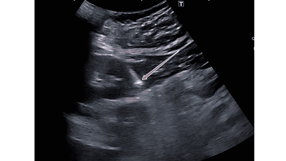
A kidney ultrasound may be ordered to look at kidney size and anatomy. Ultrasound cannot diagnose nephrotic syndrome by itself, but it can help rule out other issues and can be part of biopsy planning.
Why biopsy comes up
People are understandably nervous when a clinician mentions a kidney biopsy. The reason it comes up so often is simple: nephrotic syndrome is a pattern, not a single diagnosis. A biopsy can show what is happening inside the glomeruli and point to the specific disease, which guides treatment.
Clinicians are more likely to move toward biopsy when:
- The cause is not already clear (for example, new nephrotic syndrome without long-standing diabetes).
- Kidney function is declining or changing quickly.
- There are additional concerning findings, like significant microscopic blood, abnormal immune labs, or resistant high blood pressure.
- Treatment would involve medications with serious tradeoffs (like immunosuppressants), and the team needs the clearest diagnosis possible.
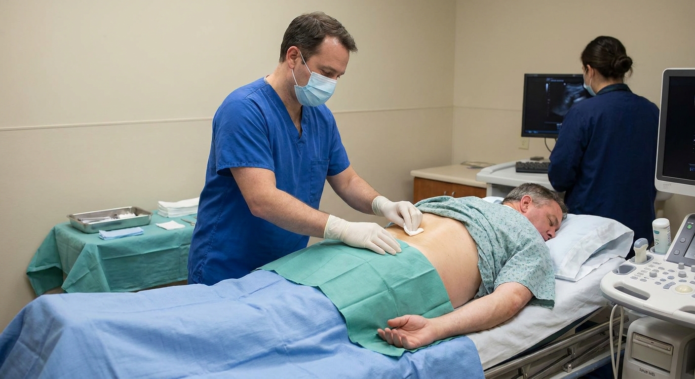
Not everyone needs a biopsy, and the decision is individualized. But at a high level, a biopsy helps clinicians choose the right medication plan instead of guessing.
Blood pressure and fluid
Even before the exact cause is nailed down, clinicians often focus on two immediate priorities: protecting the kidneys and controlling swelling.
Blood pressure control
High blood pressure can worsen kidney damage over time. Certain blood pressure medications can also reduce protein loss in the urine, which is one reason they are commonly used in proteinuric kidney disease. Many patients will hear about medications like ACE inhibitors or ARBs. In some settings, clinicians also consider SGLT2 inhibitors to help protect kidney function. Your care team will choose medications based on kidney function, potassium level, pregnancy status, and other factors.
Managing extra fluid
Swelling can be uncomfortable and, in more severe cases, can contribute to shortness of breath or skin breakdown. Fluid management may include:
- Diuretics (“water pills”) to help the body release salt and water.
- Salt restriction, because sodium tends to pull water with it.
- Daily weights at home to track fluid shifts (often more reliable than how you feel day to day).
Fluid care in nephrotic syndrome can be tricky. Too little diuretic does not help swelling. Too much can cause dizziness, kidney strain, or electrolyte problems. This is one reason close follow-up and lab monitoring matter. If you are dealing with swelling, it is also a reason not to self-adjust prescription diuretics without talking to your clinician.
Complications to know
Nephrotic syndrome is treatable, but it deserves respect. Heavy protein loss can change how the body handles fluids and clotting, and it can affect immune function. Depending on severity and cause, complications can include:
- Blood clots (venous thromboembolism), especially in more severe cases with very low albumin.
- Infections, due to urinary loss of proteins involved in immune defense and, sometimes, immunosuppressive treatment.
- Acute kidney injury or progressive kidney disease in some forms.
- Malnutrition over time if protein loss is significant and appetite is poor.
Readers often ask about blood thinners. Not everyone with nephrotic syndrome needs anticoagulation, but clinicians sometimes assess clot risk and may consider blood thinners in select high-risk situations.
If you have nephrotic syndrome, ask your clinician what warning signs should prompt urgent evaluation in your specific case.
Treatment overview
Treatment depends on the underlying cause and how severe your symptoms are. Plans usually include a mix of supportive kidney protection and cause-targeted therapy.
Supportive care
- Blood pressure management and kidney protection
- Swelling control (diuretics, salt reduction, sometimes fluid limits)
- Cholesterol management when indicated
- Vaccination review and infection risk planning when immunosuppressants are used
Cause-targeted therapy
This may involve steroids or other immune-modifying medications for certain primary kidney diseases, or targeted management of secondary causes like diabetes or lupus. The right approach is very diagnosis-specific, which circles back to why biopsy and specialized nephrology care are so often part of the process.
When to seek urgent care
Contact a clinician promptly or seek urgent evaluation if you have nephrotic syndrome (or suspect it) and notice:
- Shortness of breath, chest pain, or coughing up blood
- One leg suddenly more swollen than the other, especially with pain or warmth
- New, severe swelling with rapid weight gain over a day or two
- Fever or signs of infection
- Very little urine output or symptoms of dehydration like severe dizziness
These symptoms do not always mean a complication is happening, but they are important enough that you should not wait them out at home.
Questions to ask
- How much protein am I losing in my urine right now?
- What is my albumin level, and what does it mean for swelling and clot risk?
- Do you suspect a primary kidney disease or a secondary cause like diabetes or lupus?
- Do I need a kidney biopsy? If yes, what decision will the biopsy help you make?
- What salt and fluid targets do you want me to follow?
- What symptoms should trigger a same-day call or urgent visit?
Nephrotic syndrome can feel scary, especially when swelling appears suddenly or lab results sound unfamiliar. But with a clear diagnosis and steady follow-up, many people do very well. If you are seeing foamy urine, new swelling, or you were told you have significant protein in your urine, it is worth getting evaluated sooner rather than later.