When you are told you may need a pyeloplasty, it is completely normal to feel a little overwhelmed. In clinic, I see this often: patients understand that something is “blocked,” but the surgery itself feels like a black box. Let’s open that box together.
Pyeloplasty is a procedure that rebuilds the area where the kidney meets the ureter, called the ureteropelvic junction (UPJ), so urine can drain more easily from the kidney down to the bladder. This page focuses on what the operation and recovery tend to be like. If you are still sorting out whether your UPJ is truly blocked, your clinician will usually confirm that with imaging and functional tests before recommending surgery.
What pyeloplasty is trying to fix
Your kidney makes urine continuously. That urine collects in a central space in the kidney (the renal pelvis) and then drains into the ureter. If the UPJ is narrowed or kinked, urine can back up, potentially leading to hydronephrosis (swelling of the kidney from backed-up urine), pain, infections, or declining kidney function depending on the situation.
Pyeloplasty aims to remove the narrowed segment and reconnect healthy tissue so the pathway is wider and drains with less resistance. Think of it like replacing a pinched section of hose rather than trying to force water through the pinch.
Approaches: minimally invasive vs open
Your urologist will recommend an approach based on anatomy, prior surgeries, body size, the suspected cause of the narrowing (for example, scar tissue or a crossing blood vessel), and local expertise.
Minimally invasive pyeloplasty
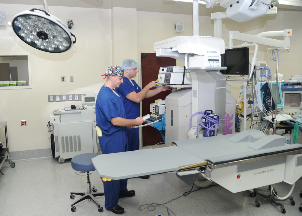
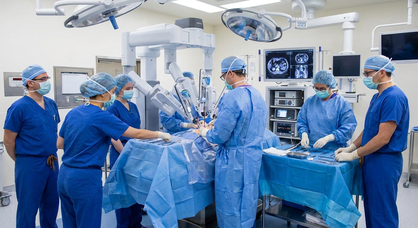
This is usually done laparoscopically (small incisions with long instruments) or robot-assisted (still small incisions, with robotic arms controlled by the surgeon). In many centers, this is the most common approach.
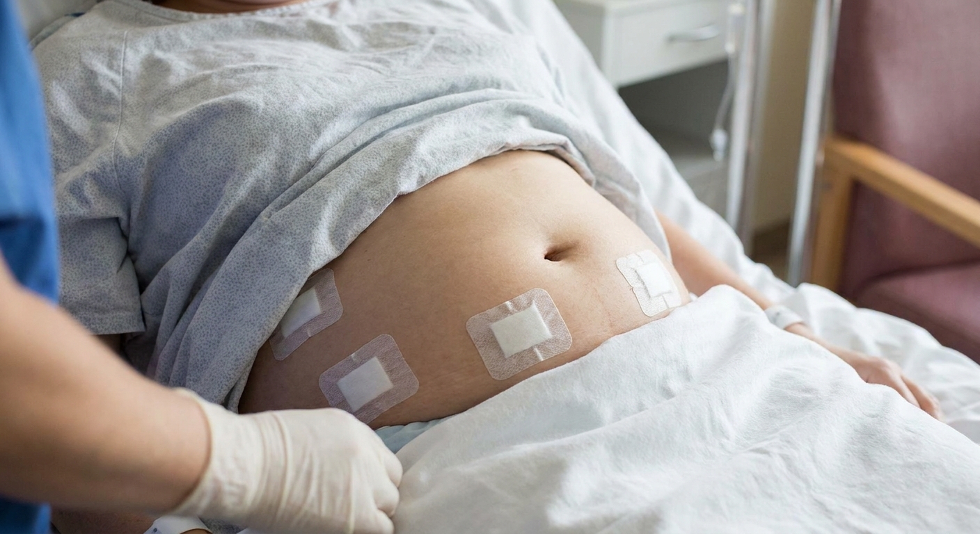
- Incisions: Several small incisions on the side or abdomen.
- Typical benefits: Less incision pain, less blood loss, shorter hospital stay, smaller scars, often a quicker return to daily activities.
- Tradeoffs: Longer operating time in some cases, and not everyone is a candidate depending on anatomy or prior surgery.
Open pyeloplasty
Open surgery uses a single larger incision (often on the flank or abdomen) to reach the UPJ directly.
- Incision: One larger incision, which usually means more soreness afterward.
- Typical benefits: Excellent exposure for complex anatomy or revisions.
- Tradeoffs: Often a longer hospital stay and longer activity restrictions.
No matter the approach, the goal is the same: a durable repair that improves drainage and protects kidney health.
Before surgery: the pre-op process
Most people have a pre-op visit or phone call where your team reviews your medical history, medications, and anesthesia plan. Common pre-op steps include:
- Medication review: You may be asked to stop certain blood thinners, anti-inflammatory medications, or supplements that increase bleeding risk. Always follow your surgeon’s instructions, and never stop prescribed blood thinners without guidance.
- Lab work and sometimes an EKG: Especially if you have other medical conditions.
- Urine testing: If there is any sign of infection, it is typically treated before surgery because operating through infection increases risk.
- Fasting instructions: These vary by facility. Many centers allow clear liquids closer to surgery but restrict solid food for a longer window. Follow the exact instructions your anesthesia team gives you.
If you have diabetes, sleep apnea, kidney disease, or a history of difficult anesthesia, bring it up early. These do not necessarily prevent surgery, but they can change the plan.
What happens during pyeloplasty
While the exact steps vary by surgeon and anatomy, most pyeloplasties include:
- Anesthesia: You will be under general anesthesia.
- Exposure of the UPJ: The surgeon reaches the kidney and upper ureter.
- Removal of the narrowed segment: The tight portion is excised.
- Reconnection (reconstruction): The renal pelvis and ureter are reattached in a way that supports better drainage.
- Temporary support for healing: Many patients have a temporary internal ureteral stent placed to keep the channel open while it heals. In selected cases, some surgeons perform a stentless repair.
In some cases, surgeons also address contributing factors, such as repositioning the connection if a crossing blood vessel is part of the problem.
Hospital stay: what to expect
Hospital length varies by country, center, and your specific situation. Many patients stay:
- Minimally invasive: sometimes same-day or 1 night, often up to 2 nights
- Open surgery: often 2 to 5 nights
Right after surgery
When you wake up, you will likely have:
- IV fluids
- Pain control (often a combination approach to minimize opioid needs)
- A urinary catheter (commonly removed within a day or so, depending on the case)
- Possibly a drain near the incision to collect extra fluid (more common in some cases than others)
It is very normal to feel groggy, sore, and bloated at first. After laparoscopic or robotic surgery, shoulder or upper chest discomfort can happen from the gas used to create working space inside the abdomen, especially with transperitoneal approaches.
The first 24 to 48 hours
Your care team typically focuses on:
- Getting you walking (even short trips in the hallway help prevent blood clots and help your bowels “wake up”)
- Advancing diet from liquids to solid food as tolerated
- Transitioning pain meds from IV to oral medicines
- Monitoring urine output and watching for fever or signs of bleeding

The stent or drain: what it is like
Many people are surprised to learn that pyeloplasty can come with temporary “hardware.” Knowing what is normal can save you a lot of worry.
Ureteral stent (internal)
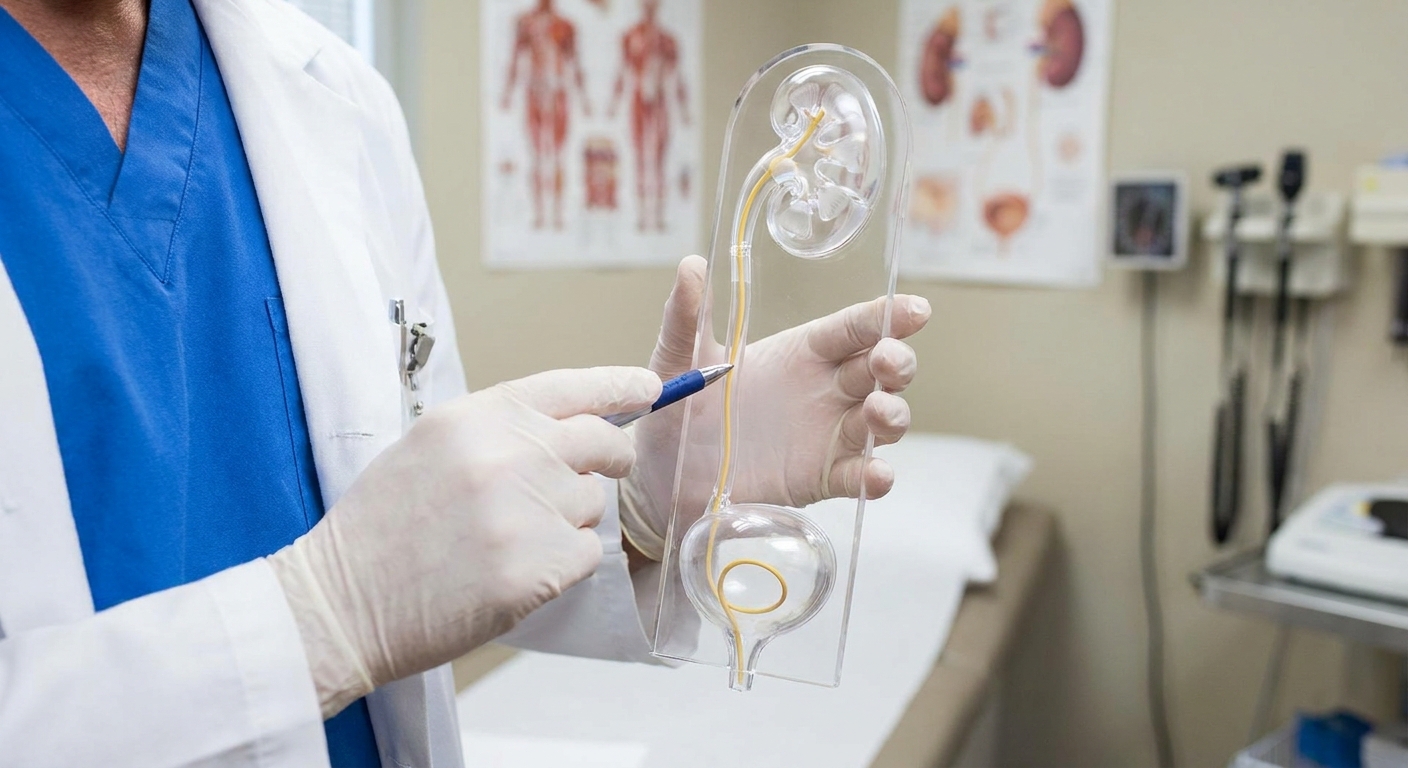
A ureteral stent is a soft tube that sits inside the ureter from the kidney area down to the bladder. It supports drainage and protects the repair while swelling goes down.
Common stent-related symptoms include:
- Urinary frequency or urgency
- Burning with urination
- Feeling like you cannot fully empty your bladder
- Blood-tinged urine, especially after activity
- Aching in the flank or bladder area, sometimes worse at the end of urination
These symptoms can be annoying, but they are often manageable. Your clinician may prescribe medications to relax the ureter or bladder, and hydration can help.
External drain (if used)
Some patients go home with a small drain, or it may be removed before discharge. The drain helps prevent fluid buildup around the repair. If you are sent home with one, your team should teach you how to measure output and what changes to report.
Recovery at home
Recovery is not just about “healing the incision.” It is about letting that reconstructed connection settle in, reducing inflammation, and gradually returning to normal activity.
Pain and fatigue
Most people feel noticeably better week by week, not day by day. Incision soreness, flank tightness, and fatigue are common early on. Many patients use a combination of:
- Acetaminophen (if safe for you)
- Prescription pain medication for breakthrough pain, if provided
- Stool softeners or gentle laxatives while on opioid medication
- Heat packs on sore muscles (avoid direct heat on incisions unless your surgeon approves)
Incision care
Follow your surgeon’s specific instructions. In general, call if you notice spreading redness, worsening swelling, pus-like drainage, or a fever.
Hydration and bladder symptoms
If you have a stent, hydration can help dilute urine and reduce irritation. Mild blood in the urine can occur, especially after activity, but large clots, inability to urinate, or heavy bleeding should be reported promptly.
Lifting and activity
Your surgeon will give the final word, but a common recovery pattern looks like this:
- First 1 to 2 weeks: focus on walking, basic self-care, and rest. Avoid strenuous chores.
- Lifting limit: many teams recommend avoiding heavier lifting early on (often around 10 to 15 pounds), with longer restrictions after open surgery. Your instructions may be different depending on your incision and repair.
- Driving: typically once you are off opioid pain medications and can comfortably brake and turn.
- Work: desk work may be possible sooner than jobs that require heavy lifting, bending, or long hours on your feet.
If you are unsure whether something counts as “too much,” my practical rule is this: if an activity increases flank pain, stent discomfort, or causes noticeable bleeding in the urine afterward, scale back and check in with your team.

Timeline snapshot
Every surgeon has their own protocol, but a very common arc looks like this:
- Week 1: walking, pain control, and getting your appetite and bowels back on track
- Weeks 2 to 4: gradual return to routine daily activities, still avoiding heavy lifting
- Around 2 to 8 weeks: stent removal if you have one (many are removed around 4 to 6 weeks)
- Around 6 to 12 weeks or later: follow-up imaging in many protocols, sometimes later depending on symptoms and surgeon preference
When is the stent removed?
Stent timing varies. A very common range is about 4 to 6 weeks, but some are removed earlier or later depending on the repair, symptoms, and surgeon preference. Removal may be done in clinic with a quick procedure (often cystoscopy). Many patients describe it as uncomfortable but brief.
After stent removal, some people have a day or two of urinary burning, frequency, or mild flank aching. Your clinician will tell you what to expect and when to call if symptoms feel out of proportion.
Follow-up imaging
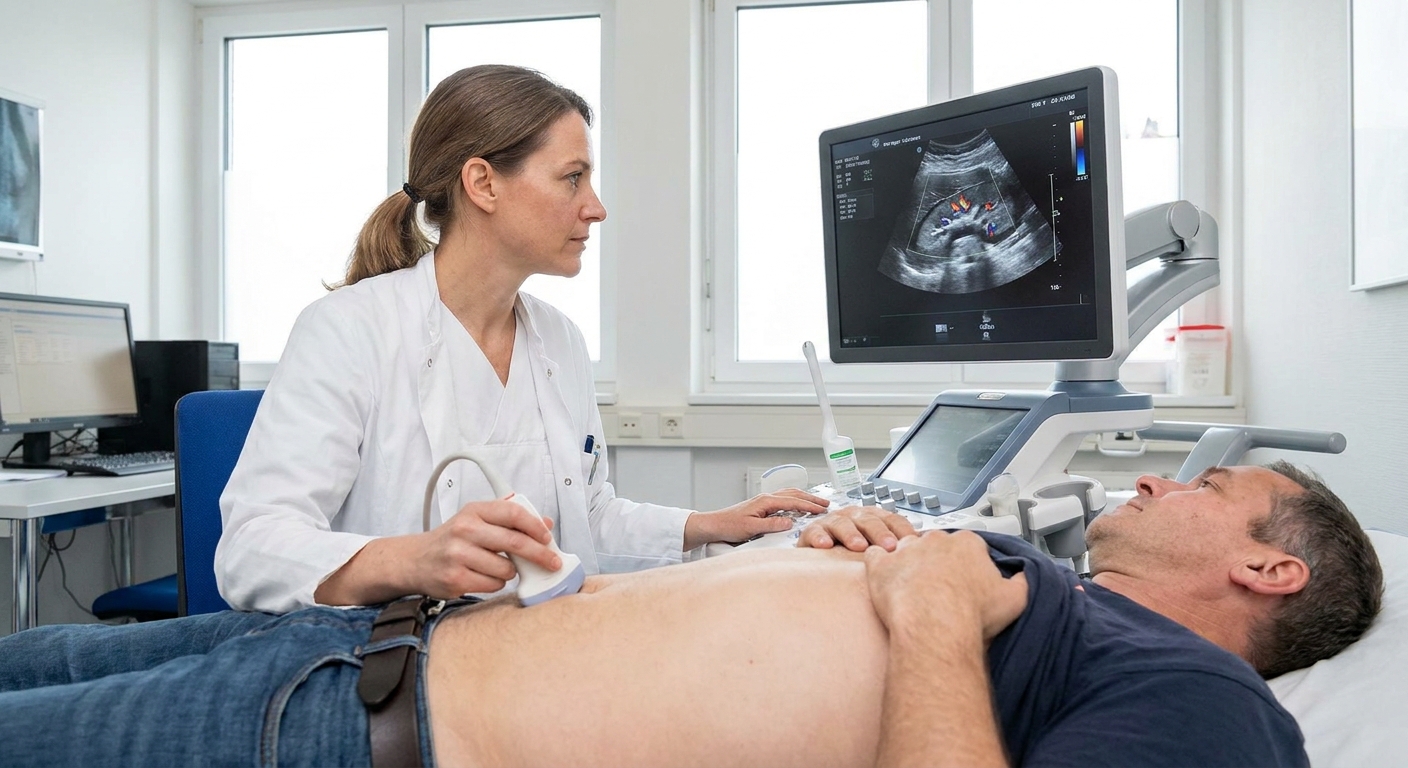
A successful pyeloplasty is judged by symptoms, kidney health, and how urine drains. It is very common to have a follow-up scan after you have healed to assess drainage and kidney function. Depending on your situation and local practice, your clinician may use tests such as:
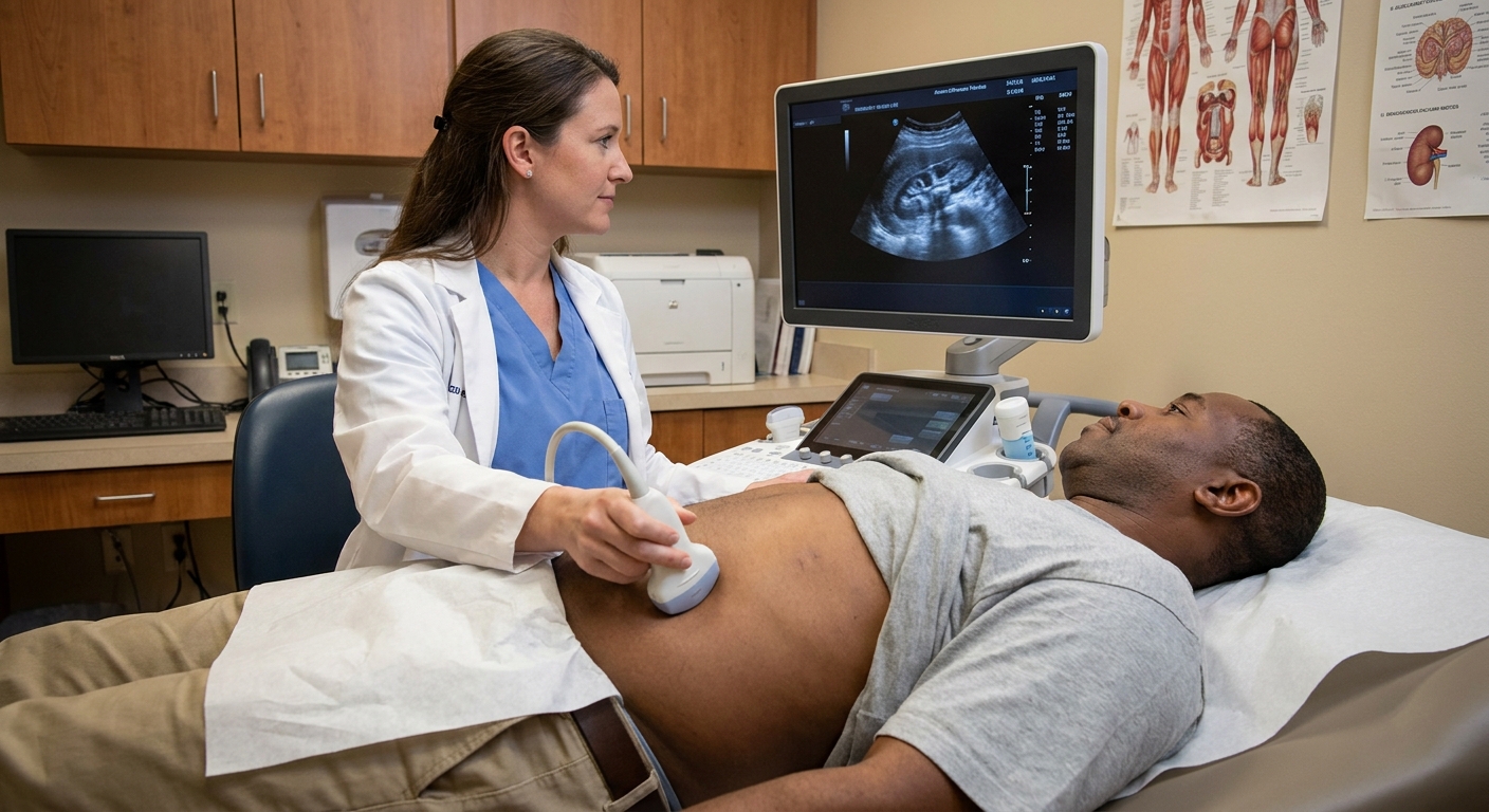
- Renal ultrasound to look at hydronephrosis over time
- Nuclear medicine renal scan (often called a diuretic renogram) to evaluate drainage dynamics and split kidney function
- CT or MRI in selected cases
A key point I share with patients: hydronephrosis can improve slowly. Some swelling may persist even when drainage is better, so your clinician interprets imaging in context.
Success rates and what “success” means
In experienced hands, pyeloplasty has high success rates. Success usually means improved drainage on follow-up testing, stable or improved kidney function, and fewer symptoms like pain or infections. Your surgeon is the best person to quote success rates for your anatomy, approach, and whether this is a first-time repair or a revision.
Alternatives in some cases
Not everyone needs immediate reconstruction, and not every UPJ problem is treated the same way. Depending on symptoms, kidney function, and imaging, your clinician may discuss options such as:
- Observation with follow-up imaging in select cases, especially if symptoms are minimal and kidney function is stable
- Temporary drainage (for example, a ureteral stent or nephrostomy tube) if urgent decompression is needed
- Endoscopic options such as balloon dilation or endopyelotomy in selected patients (these are not right for everyone, and durability can differ from pyeloplasty)
If alternatives come up in your visit, ask what makes you a good candidate or not, and how your team defines a durable result.
Possible risks and complications
All surgeries have risks, and your surgeon will review these in detail for your specific case. In plain language, potential concerns include:
- Infection (urinary or at the incision)
- Bleeding
- Urine leak from the repair site (sometimes requiring longer drainage or additional procedures)
- Scar tissue or recurrent narrowing over time
- Injury to nearby structures (uncommon, but part of standard informed consent)
- Stent-related issues such as discomfort, migration, or rarely blockage
Most people do very well, especially when they are careful about recovery instructions and keep follow-up appointments. Still, I never want patients to feel blindsided, so it is worth knowing what you are watching for.
Call your surgeon or seek urgent care if
After pyeloplasty, do not “tough it out” through symptoms that could signal a complication. Contact your surgical team urgently or seek emergency care if you have:
- Fever (especially 100.4°F or 38°C or higher) or chills
- Worsening flank pain that is not controlled with your prescribed plan
- Inability to urinate
- Heavy bleeding in the urine, large clots, or urine that looks like dark red wine
- Persistent vomiting or inability to keep fluids down
- Shortness of breath, chest pain, or one-sided leg swelling
- Increasing redness, warmth, or pus-like drainage from an incision
Questions to ask pre-op
- Which approach are you recommending for me and why?
- How long do you expect I will be in the hospital?
- Will I have a ureteral stent, an external drain, or both?
- When will the stent come out, and how is it removed?
- What are my lifting limits, and for how long?
- When can I drive, return to work, and exercise?
- What follow-up imaging will I need, and when?
- What symptoms are normal with the stent, and what symptoms are not?
Bottom line
Pyeloplasty is a reconstructive surgery designed to improve urine drainage from the kidney by repairing the ureteropelvic junction. Whether your procedure is minimally invasive or open, most patients can expect a short hospital stay (with real variability by center), possible temporary stenting, activity restrictions especially around lifting, and follow-up imaging later to confirm the kidney is draining well.
If you are preparing for surgery, bring your questions. You deserve to understand what is happening inside your body and what a safe, steady recovery should look like.