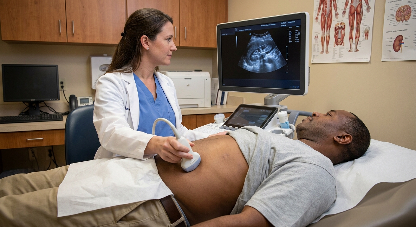
I still remember the first time a patient brought me an ultrasound report that said “possible hydronephrosis.” They were already bracing for the worst, picturing a blocked kidney and an urgent procedure. But when we slowed down and looked at the wording and follow-up imaging, the “backup” turned out to be something entirely different: a parapelvic cyst. Same neighborhood in the kidney, very different meaning.
If your report mentions a “parapelvic cyst,” “peripelvic cyst,” “renal sinus cyst,” or “hydronephrosis vs parapelvic cyst,” this article is for you. I will walk you through what each term means, why they get confused, and what radiologists do to separate a fluid-filled cyst from true urine backup in the collecting system.
First, the anatomy
Your kidney has a urine collecting system, called the renal pelvis and calyces, that funnels urine into the ureter and down to the bladder. Hydronephrosis means that collecting system is dilated because urine is not draining normally.
A parapelvic cyst is a fluid-filled sac that sits beside the renal pelvis, often near or within the renal sinus region. It is not part of the collecting system. It is usually benign and usually not an obstruction by itself.
You may also see the term peripelvic cyst. In radiology, people sometimes use parapelvic and peripelvic loosely in conversation, but they can refer to slightly different origins and locations. Parapelvic cysts are often thought of as simple renal cysts that extend toward the renal sinus, while peripelvic cysts are more classically described as cysts within the renal sinus, sometimes related to lymphatic channels. On imaging reports for patients, both may be summarized as a “renal sinus cyst,” and either can mimic hydronephrosis depending on size and location.
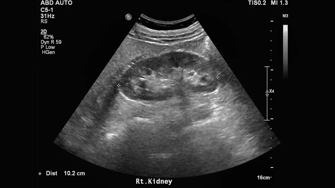
The tricky part is that both are fluid, and both live in the center of the kidney where the pelvis and calyces sit. On a single still image, especially on ultrasound, they can mimic one another.
How they look on imaging
Hydronephrosis: connected branching fluid
Radiologists look for a connected, branching pattern that matches the calyces and renal pelvis. The dilated calyces often look like little extensions reaching outward from the pelvis. Another key clue is that the fluid spaces tend to look like one continuous system rather than separate bubbles.
You might also see softer wording like “pelviectasis” or “mild fullness”. Those terms often imply mild dilation and can be transient or non-obstructive, but they still need to be interpreted in context.
Parapelvic cyst: rounded pockets that do not connect
Parapelvic (and peripelvic) cysts typically appear as well-defined, more rounded fluid collections. They may be single or multiple. When there are several, they can cluster near the pelvis and create an overall hydronephrosis-like impression, but the individual pockets often look separate rather than truly branching into calyces.

How ultrasound helps sort it out
Ultrasound is often the first test that raises the question, and it is also where the confusion most commonly happens. When the answer is not clear on initial images, radiologists lean on technique and a few specific tells.
1) Extra angles and sweeps through the renal sinus
A major practical difference is continuity. Sonographers and radiologists will sweep through the kidney in multiple planes to see whether the fluid spaces merge into a single collecting system (more consistent with hydronephrosis) or remain discrete rounded spaces (more consistent with cysts).
2) Looking for the ureter and downstream clues
If hydronephrosis is real and due to obstruction, there may be supportive findings such as:
- Hydroureter (a dilated ureter) in some causes of obstruction
- Bladder abnormalities or high post-void residual in certain settings
- A visible stone with acoustic shadowing, although many stones are not seen on ultrasound
Parapelvic or peripelvic cysts do not drain into a ureter, so you do not typically see a dilated ureter as a direct consequence of the cyst.
3) Color Doppler to avoid a common pitfall
Radiologists also use Doppler to make sure a suspected fluid space is not actually a blood vessel near the renal hilum. Doppler is not what distinguishes urine from cyst fluid, but it helps avoid mislabeling vascular structures as hydronephrosis.
4) A very common mimic: extrarenal pelvis
Another reason a report may hedge is an extrarenal pelvis, which is a normal anatomic variant where part of the renal pelvis sits outside the kidney. On ultrasound it can look like mild hydronephrosis, but it lacks the branching calyceal dilation you would expect with true obstruction.
5) When ultrasound cannot decide
Sometimes ultrasound can only say, honestly, “hydronephrosis versus parapelvic cysts.” That is not a failure. It is a cue that a different modality, often CT or MRI, may be the best next step depending on your symptoms, pregnancy status, and kidney function.
How CT settles the question
When radiologists need to be sure whether the collecting system is actually dilated, contrast-enhanced CT with delayed (excretory) images can be extremely helpful. Two important caveats: not every CT is performed with an excretory phase unless it is specifically ordered, and severe obstruction can delay or reduce contrast excretion from the affected kidney.
The key idea: excreted contrast outlines urine pathways
After IV contrast is given, the kidneys filter it and excrete it into the urine. On delayed (excretory) phase images, the contrast is visible within the renal pelvis, calyces, and ureters.
This is often the most definitive imaging clue for the collecting system:
- If it is hydronephrosis, the dilated spaces are part of the collecting system, so they will fill with contrast-opacified urine on delayed images (unless excretion is markedly delayed).
- If it is a parapelvic or peripelvic cyst, it will not fill with contrast. It stays a separate, non-enhancing fluid collection adjacent to the contrast-filled collecting system.
Radiologists also look for a cause of obstruction on CT, such as a stone, mass effect, or stricture, and for secondary signs like perinephric stranding. Perinephric stranding can be seen with acute obstruction, but it is nonspecific and can also occur with infection or other inflammation.
What about MRI and renal scans?
MRI urography
MRI can also differentiate cysts from collecting system dilation. It can do this in more than one way:
- Heavily T2-weighted (noncontrast) MR urography makes fluid stand out and can help map whether fluid spaces connect like a collecting system.
- Contrast-enhanced MR urography can add excretory information similar to CT, but gadolinium contrast still has kidney-function considerations in some patients.
MRI is sometimes chosen when radiation avoidance is important, including in pregnancy, or when CT is not ideal for other clinical reasons.
Renal scan (diuretic renography)
When the question is less “is this a cyst?” and more “is drainage impaired?”, a nuclear medicine renal scan can evaluate kidney function and how well urine drains, sometimes with a diuretic challenge. This can be useful when hydronephrosis is present but the degree of obstruction is uncertain.
How doctors decide what happens next
In real life, the plan is usually driven by two things: your symptoms and labs, and how confident the imaging is.
- Mild findings, no symptoms, stable kidney function: often conservative management, sometimes no follow-up if the imaging is classic for a benign cyst.
- Unclear ultrasound: clarification with CT (often with an excretory phase when appropriate) or MRI is common.
- Red flags such as significant pain, fever, rising creatinine, a solitary kidney, pregnancy with concerning symptoms, or suspected infected obstruction: this is treated as urgent until obstruction and infection are addressed.
Why the wording matters
This distinction is not just academic. A report that calls something hydronephrosis can set off a chain of events: repeat imaging, urology referral, lab work, and sometimes urgent evaluation if symptoms suggest acute obstruction.
When radiology can confidently call the finding a parapelvic or peripelvic cyst, it often prevents an obstruction workup that is unlikely to help, especially if:
- You have no flank pain, fever, or urinary symptoms
- Your kidney function labs are stable
- There is no evidence of a blocked ureter on follow-up imaging
That said, these cysts can occasionally be associated with symptoms or coexist with other issues, so your clinician still interprets the imaging in the context of you, not just the picture.
What a parapelvic cyst means for you
Most parapelvic and peripelvic cysts are benign incidental findings. They may be found during evaluation for unrelated abdominal pain, blood in the urine, kidney stones, or a screening ultrasound.
Common next steps depend on the situation:
- No symptoms and classic imaging appearance: often reassurance, sometimes no follow-up needed.
- Unclear appearance on ultrasound: CT or MRI may be recommended to clarify.
- Symptoms that suggest obstruction or infection: your clinician may treat this as urgent until obstruction is confidently excluded.
If your report mentions parapelvic or peripelvic cysts and you have ongoing pain, recurrent urinary tract infections, or blood in your urine, it is reasonable to ask whether the cyst could be contributing or whether something else is going on alongside it.
Questions to ask
- Does the report say hydronephrosis, possible hydronephrosis, or hydronephrosis vs parapelvic cyst?
- Was this based on ultrasound alone, or do I have delayed contrast images (excretory phase) that evaluate the collecting system?
- Is there any mention of a dilated ureter or a suspected cause like a stone?
- Do my symptoms or labs suggest obstruction, infection, or stone disease that needs attention?
- Should I repeat imaging, and if so, what test is most appropriate for me based on kidney function, pregnancy status, and radiation or contrast considerations?
When to seek urgent care
Even though parapelvic and peripelvic cysts are usually harmless, do not ignore symptoms that could signal a true obstruction or infection. Seek urgent evaluation if you have:
- Fever, chills, or feel severely ill
- Moderate to severe flank pain, especially with nausea or vomiting
- New trouble urinating or very low urine output
- Known kidney disease with worsening symptoms
- Severe pain plus blood in the urine
These symptoms can occur with an obstructing stone or kidney infection, and those need prompt care regardless of what a prior incidental cyst showed.
FAQ
Can a parapelvic cyst cause hydronephrosis?
It is uncommon, but a large cyst or multiple cysts could potentially compress the collecting system or the ureteropelvic junction and contribute to obstruction. More often, the issue is simply that parapelvic or peripelvic cysts look like hydronephrosis on certain images. If there is concern for true obstruction, delayed excretory imaging or functional testing can help clarify.
Why did my ultrasound say hydronephrosis, but my CT said parapelvic cyst?
Ultrasound is excellent, but it can have limitations depending on body habitus, bowel gas, and viewing angle. CT can more definitively show whether the fluid is within the collecting system (hydronephrosis) or separate (cyst), especially when delayed excretory images are included.
Does a parapelvic cyst need treatment?
Most do not. Management is individualized and depends on symptoms, size, and whether there are complications or another diagnosis that better explains what you are feeling.
What words in a report suggest a cyst instead of obstruction?
Phrases like “parapelvic cyst,” “peripelvic cyst,” “renal sinus cyst,” “no calyceal dilatation,” “no hydroureter,” “extrarenal pelvis,” or “collecting system not dilated on delayed images” generally point away from a clinically significant obstruction. Your clinician can interpret the exact wording with you.
The bottom line
Hydronephrosis means urine backup in the kidney’s collecting system and can signal a blockage that needs timely evaluation. Parapelvic and peripelvic cysts are separate fluid pockets near the renal pelvis that can mimic hydronephrosis, especially on ultrasound. Another common mimic is an extrarenal pelvis.
Radiologists sort this out by looking for continuity and a branching collecting system, and when needed, by using CT or MRI techniques that evaluate the urine pathways. When the finding is truly a cyst, calling it correctly can spare you the stress and testing that comes with an “obstruction” label.