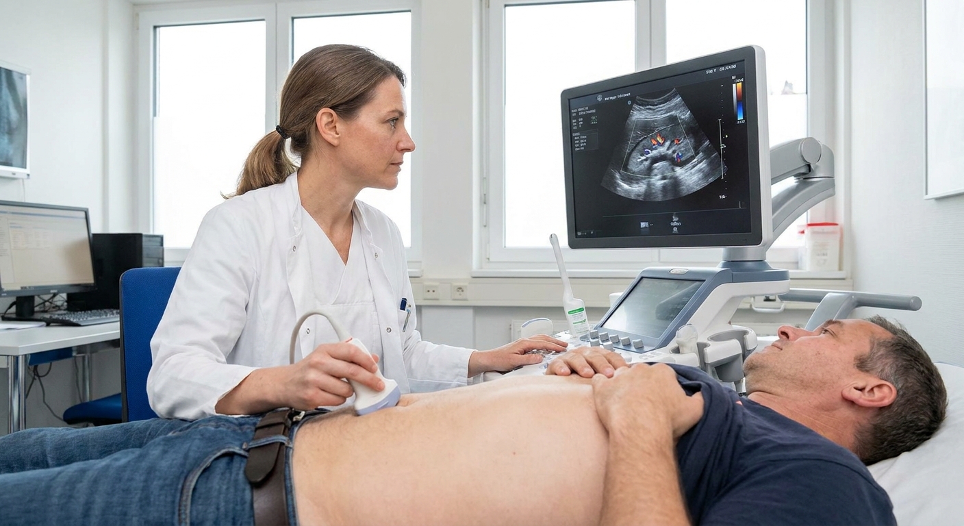
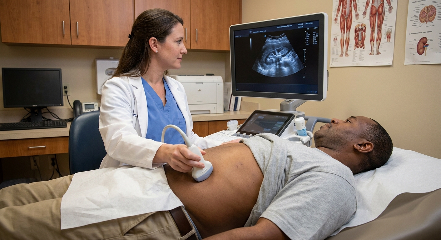
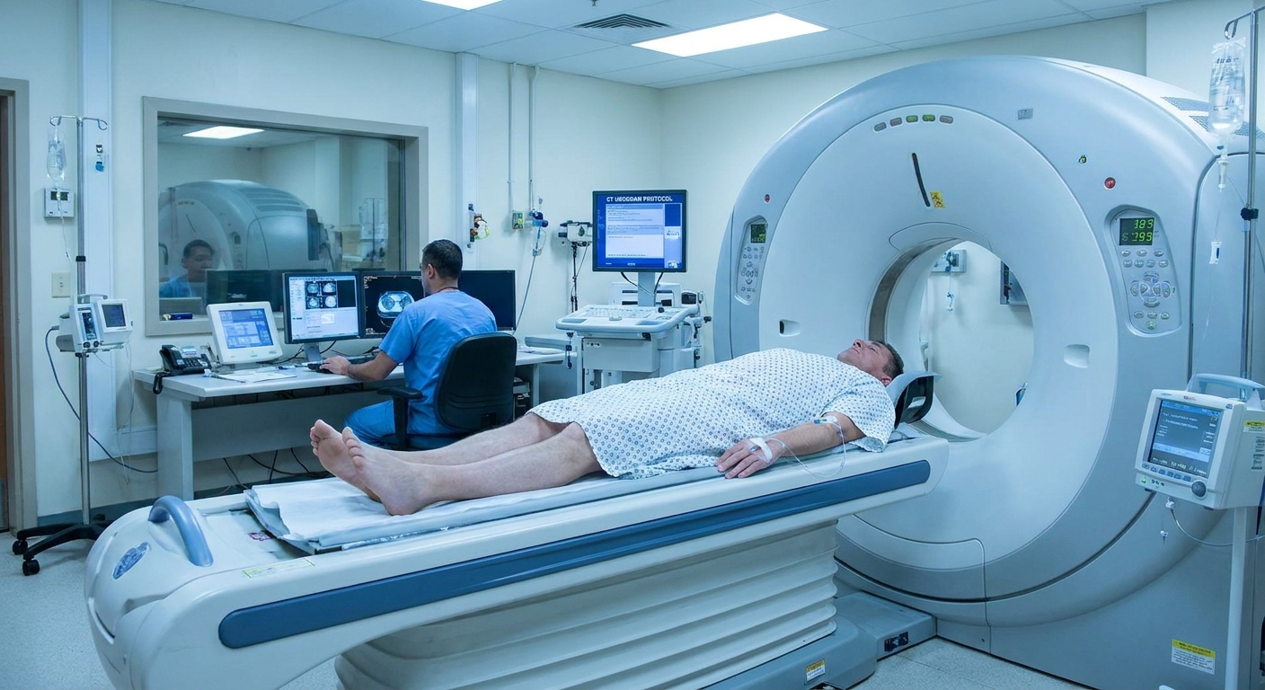
When I worked in a busy family clinic, one of the most common frustrations I heard was, “No one told me what the appointment would actually be like.” A MAG3 renal scan is a great example. The name sounds intimidating, and the word nuclear can make anyone uneasy. In reality, most people describe it as more boring than scary. You lie on a table while a camera takes pictures of how your kidneys take up the tracer and how urine drains, and at some point you may receive a medication that makes you urinate more.
This page walks you through a MAG3 renal scan in plain language so you can show up knowing what to expect. In many cases, a MAG3 scan is done as a diuretic renogram, which is a specific protocol that adds a diuretic medication to better assess drainage.
Why your clinician ordered it
A MAG3 renal scan is usually ordered to answer very practical questions that other tests cannot always settle. Common reasons include:
- Possible blockage or narrowing (for example, suspected UPJ obstruction) when ultrasound or CT shows hydronephrosis and the next question is whether drainage is actually impaired
- Measuring split kidney function (how much the right kidney contributes vs the left) when that number affects treatment decisions
- Follow-up after surgery on the kidney or ureter, to see how drainage looks afterward
- Evaluation of kidney drainage patterns in people with recurrent infections, pain episodes, or complicated urinary tract anatomy
Your situation matters. Two people can have the same ultrasound report but need different next steps, and this scan is often used to add that missing “how well does it actually work” piece.
What it measures
A MAG3 renal scan uses a tiny amount of a radioactive tracer (a substance that gives off a signal the camera can detect). The tracer travels through your bloodstream to your kidneys. Your kidneys remove it from the blood (mostly through tubular secretion) and pass it into urine.
As the tracer moves, a special camera takes rapid images over time. This lets the nuclear medicine team answer two practical questions:
- How well is each kidney functioning? This is often reported as a “split” or “differential” function, meaning the percentage of total kidney function contributed by the right vs left kidney.
- How well does urine drain from the kidneys? This is evaluated by watching how quickly the tracer leaves the kidney area and moves downstream.
Many MAG3 scans focused on drainage are done as a diuretic renogram, meaning a diuretic medication is given during the test to encourage urine flow and help clarify whether urine is draining freely.
Before you arrive
Your imaging center will give instructions that match your situation, but these are the most common preparation steps:
- Hydration matters. Many facilities want you well hydrated so the test is easier to interpret and you can urinate afterward. Some will ask you to drink water before the scan, while others give fluids through an IV.
- Ask about your medicines. Do not stop prescriptions unless the ordering clinician or imaging center specifically tells you to. Some centers give special instructions for certain diuretics or blood pressure medicines.
- Plan to be there 1 to 2 hours. Many protocols fit in this window, but timing varies. If you are told it is a “diuretic renogram,” budget extra time and ask your lab what they typically schedule.
- Wear comfortable clothing. You may be asked to change into a gown and remove metal from the waist area.
If you are pregnant, might be pregnant, or are breastfeeding, tell the imaging team before the tracer is given. The plan can change depending on your specific circumstances.
What happens during the scan
1) Check-in and a quick screening
You will be asked about allergies, current medications, kidney history, recent imaging, and for anyone of childbearing potential, pregnancy status. This is also the right moment to speak up about:
- difficulty lying flat
- claustrophobia
- back pain
- trouble holding urine
- a history of fainting with IVs
2) An IV is placed
A technologist places an IV, usually in your arm or hand. This IV is used to inject the MAG3 tracer and, in many protocols, the diuretic medication later in the scan.
The IV placement feels like any other blood draw or IV start: a quick pinch, then pressure.
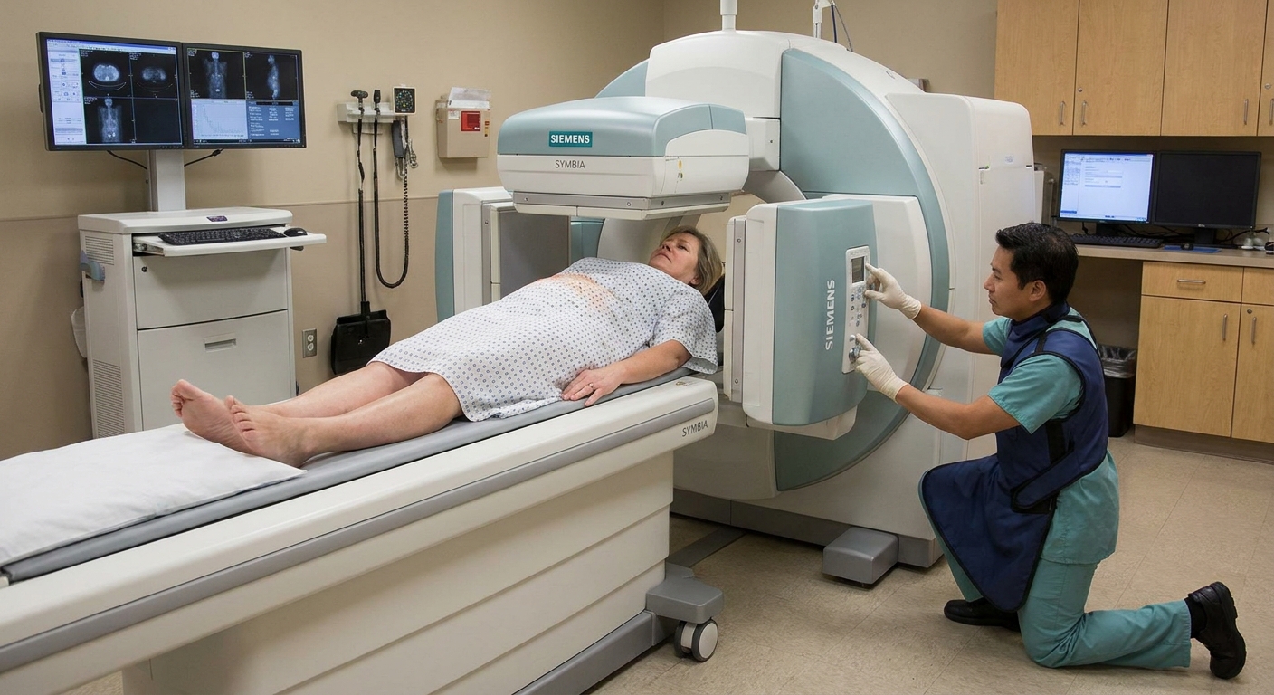
3) You lie on the table and the camera records images
You will lie on a padded table, typically on your back. The camera detector is large and comes close to your body. In many labs, one detector is positioned above your abdomen, and sometimes a second detector sits behind your back to collect images from both sides. It does not touch you, and the scan is not painful.
Once imaging begins, the technologist injects the tracer through the IV. The camera then takes a series of images over time to watch:
- how quickly tracer reaches each kidney (blood flow and uptake)
- how well each kidney processes it
- how tracer moves from kidney into the collecting system and down the urinary tract
Your job during this part is mostly to lie as still as you reasonably can. Motion can blur the data and make the curves harder to interpret. If you need to cough, reposition, or take a break, tell the technologist rather than forcing yourself to stay rigid.
If you cannot hold urine during the scan, say so immediately. This happens. The team can usually pause, help you get to the bathroom, and adjust the plan so the study is still useful.
The diuretic part
In a diuretic renogram, you will receive a diuretic medication, most commonly furosemide (Lasix). This medication encourages your kidneys to make more urine temporarily.
The point is not to “treat” anything in that moment. The point is to create a stronger flow of urine so the scan can better assess drainage under a gentle stress test.
When is it given?
There are different protocols, and your order may specify one. Common timing includes:
- Diuretic after the tracer has had time to collect in the kidney’s drainage system
- Diuretic at the beginning or even slightly before tracer in select protocols
The technologist will follow a standardized method used by that lab and the ordering clinician.
What does it feel like?
Many people feel nothing at the moment it is injected. Within minutes, you may notice:
- a strong urge to urinate
- more frequent urination
- mild lightheadedness, especially if you are sensitive to diuretics, your blood pressure runs low, or you were not well hydrated
Tell the team right away if you feel dizzy, nauseated, short of breath, develop hives, or feel unwell. Side effects are usually mild and temporary, but you deserve prompt attention if something does not feel right.
Will I need a catheter?
Sometimes, but not always. Many adult outpatient diuretic renograms are done without a catheter. A bladder catheter is a small tube placed into the bladder to continuously drain urine during the test.
A catheter may be used when:
- accurate drainage measurement is especially important
- there is concern you may not be able to empty your bladder well (for example, significant urinary retention or neurogenic bladder)
- a very full bladder could “back up” pressure and make drainage look worse than it truly is
- your clinician wants the most controlled conditions possible
If a catheter is planned, the team should explain it beforehand. Catheter placement can be uncomfortable, and it can feel personal and vulnerable. If you have a history of trauma, severe anxiety, or prior catheter difficulties, mention that up front so the team can take a more supportive approach and talk through options.
Understanding the curve
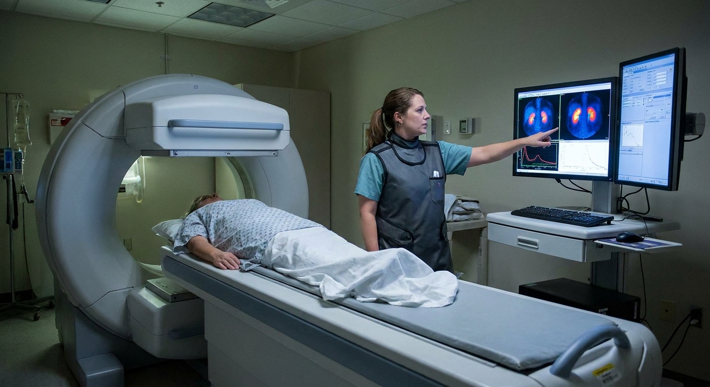
During the scan, the computer creates a time-activity graph, often called a renogram curve. One part people hear about a lot is the washout after the diuretic.
Here is the simple way to picture it:
- Tracer arrives at the kidney, so the line on the graph goes up.
- The kidney processes tracer into urine, so the kidney region shows activity.
- Tracer drains away through the kidney’s collecting system and out, so the line should go down as the tracer “washes out.”
Fast vs slow washout
- Fast washout generally suggests urine is draining freely when urine flow is encouraged.
- Slow washout can suggest delayed drainage. This does not automatically mean there is a fixed blockage. It can also be influenced by hydration, bladder fullness, overall kidney function, and anatomy.
You may also hear a term like T½ (half-time), which is an estimate of how long it takes for activity to fall by about half during the washout phase. How it is calculated can vary by lab and protocol, and it is one tool among several, not a standalone verdict.
If you take only one thing from this section: the MAG3 scan is less like a single snapshot and more like a short movie. The “curve” is a way of summarizing how that movie looks over time.
After the scan
You will likely empty your bladder
Many labs have you urinate near the end of the scan, and sometimes they take an extra set of images after you empty your bladder. This helps them see how bladder fullness may have affected drainage.
Drink fluids
If you received a diuretic, you may urinate more for a few hours. Drinking water afterward can help you feel better and helps your body clear the tracer.
When will I get results?
A nuclear medicine physician or radiologist interprets the study, then your ordering clinician reviews it with your bigger medical picture. Timing varies, but many results are available within a few business days. If you are waiting and feeling anxious, it is reasonable to ask the imaging center when reports are typically finalized.
Safety and common concerns
Is the radiation dangerous?
The tracer dose used for a MAG3 renal scan is small. Like any medical radiation exposure, it should be used only when the potential benefit outweighs the risk. For most people, the risk from this test is considered low, and the information it provides can be very valuable for decision-making.
In everyday life, most people can be around others as usual afterward. If you are breastfeeding, follow the specific guidance given by your imaging facility for your tracer and your situation.
Will I feel the tracer?
No. The tracer is not something you can feel. What you may feel is the IV placement and, if used, the diuretic effect.
Is it safe with kidney disease?
MAG3 is often chosen specifically because it is well suited for evaluating kidney function and drainage, including when function is reduced. That said, the ordering clinician and nuclear medicine team consider your overall kidney status when selecting a protocol.
What if I have anxiety or claustrophobia?
This test is usually done in an open room with the camera close to you, not in a tunnel like an MRI. Still, anxiety is real. Ask ahead of time if you can:
- bring a support person to the department (policies vary)
- listen to music (some labs allow it)
- use breathing techniques or grounding exercises
- discuss medication options with your prescribing clinician if your anxiety is severe
Kids and parents
For children, the overall idea is the same, but the experience can feel different. Some pediatric labs:
- encourage a parent to be present (policies vary)
- use a catheter more often to control bladder emptying and improve accuracy in small bladders
- focus heavily on comfort strategies to help a child hold still
If you are bringing a child, ask the department what you can do to help them stay calm and still, and whether there are any special hydration instructions.
When to call after
Most people have no issues afterward. Contact your clinician if you develop:
- fever, chills, or burning with urination (could suggest a urinary infection, especially if a catheter was used)
- new or worsening flank pain
- persistent vomiting or inability to keep fluids down
- significant dizziness, fainting, or symptoms of dehydration
If you have severe symptoms, seek urgent care.
Questions to ask
- Is my scan a standard MAG3 renal scan or a diuretic renogram?
- Should I drink water beforehand? If so, how much and when?
- Should I take my usual medications the morning of the test?
- Will a bladder catheter be used for my scan?
- About how long will I be in the department?
- When and how will my results be shared?
As a clinician, I can tell you this: the people who ask these questions are not being “difficult.” They are being prepared. And prepared patients usually have a smoother experience.

FAQ
How long does a MAG3 renal scan take?
Many scans take about 60 to 90 minutes. Some protocols are shorter, and some can run closer to 2 hours, especially if extra images are needed. Your best estimate comes from the specific lab performing your test.
Do I have to fast?
Often, no. Some centers may give specific instructions. Follow the guidance from your imaging facility because preparation can differ.
Can I drive myself home afterward?
Most people can drive themselves. If you are given medication for anxiety or you are prone to lightheadedness with diuretics, ask your clinician whether you should arrange a ride.
Will the scan tell me if I have UPJ obstruction?
The scan provides important information about function and drainage, including the washout pattern after a diuretic. Whether that pattern represents a true obstruction is a medical interpretation that depends on your symptoms, other imaging, the protocol used, and the complete report from the interpreting physician and your urology or nephrology team.