In clinic, one of the most confusing phrases people hear after an ER visit or a scan is, “There’s a narrowing in the ureter.” If you are in pain, nauseated, and worried about your kidneys, it can be hard to understand what that actually means. A ureteral stricture is one possible cause of a kidney that looks swollen on imaging, and it is different from a stone that is still on the move.
Let’s walk through what a ureteral stricture is, what symptoms it can cause, how it shows up on imaging, and what treatment usually looks like.
What a ureteral stricture is
Your kidneys make urine, which drains down thin tubes called ureters into the bladder. A ureteral stricture is a fixed narrowing in a segment of the ureter, most often caused by scar tissue.
Think of the ureter like a soft, flexible straw. If part of that straw heals with stiff scarring, the inner channel becomes smaller. When urine cannot drain easily, pressure backs up toward the kidney. That back-pressure can lead to hydronephrosis, which is swelling and stretching of the kidney’s collecting system.
Common causes
Most strictures are acquired, meaning you were not born with them. They tend to show up after something inflamed or injured the ureter and healing left behind scar tissue.
After a stone
A stone can irritate the ureter’s lining as it passes. Less commonly, a stone that was stuck for a while can inflame the wall enough that the healing process later narrows the ureter at that spot, even after the stone is gone.
After a procedure
Strictures can form after procedures that involve the ureter, such as ureteroscopy for stones, stent placement, or other endoscopic work. This does not mean the procedure was “done wrong.” Even with excellent technique, tissue can scar as it heals.
After pelvic or abdominal surgery
Surgeries near the ureter can lead to narrowing later due to inflammation, accidental injury, or scarring where the ureter was handled or reimplanted.
After radiation
Radiation therapy in the pelvis or abdomen can cause delayed scarring and narrowing of ureter tissue months to years later.
Less common causes
- Inflammatory conditions that affect the retroperitoneum or urinary tract
- Tumor within the ureter (urothelial cancer) or compression from outside the ureter (this is not always a true scar stricture, but it can look similar on imaging)
- Retroperitoneal fibrosis that traps or kinks the ureter
- Injury from trauma
Symptoms
Symptoms vary based on how tight the narrowing is, where it is located, whether it affects one or both ureters, and whether the other kidney can pick up the slack.
Flank pain
Many people describe a deep ache or pressure in the flank, meaning the back and side under the ribs. It may be intermittent or persistent. It can worsen when urine flow increases, such as after drinking a lot of fluids or anything that causes diuresis.
Urinary tract infections
When urine does not drain normally, bacteria have more opportunity to grow. Some people with strictures develop recurrent UTIs, sometimes with fever.
Nausea, vomiting, and feeling unwell
These can happen with significant obstruction, especially if pain is severe or infection is present.
A rise in creatinine
This is an important point that often gets misunderstood. If a stricture blocks one ureter and your other kidney is healthy, your blood creatinine can stay normal because the other kidney compensates.
Creatinine is more likely to rise when:
- There is only one functioning kidney
- Both kidneys are affected (for example, strictures on both sides, or a stricture plus another issue on the other side)
- The other kidney already has reduced function from chronic kidney disease
- The obstruction is severe or prolonged
That is why some people can have significant hydronephrosis on imaging and still have normal labs, while others see a quick change in creatinine.

How it shows up on imaging
Imaging is usually what raises the suspicion for a stricture, especially if hydronephrosis is present. A common pathway is an initial ultrasound or CT, followed by more targeted testing (CT urogram, MR urogram, functional testing, or a urology study) depending on what the first scan shows and what your clinician is trying to answer.
One key nuance is worth stating clearly: hydronephrosis does not always equal clinically significant obstruction. Sometimes it reflects a transient or partial blockage, reflux, pregnancy-related physiologic dilation, or other functional causes. That is why follow-up imaging or functional testing can matter.
Ultrasound
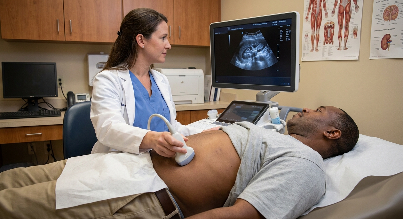
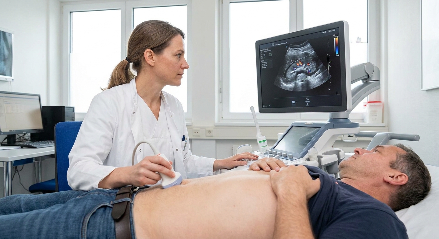
Ultrasound is great for identifying hydronephrosis without radiation. It may not always show the ureter clearly or pinpoint the exact level of narrowing, but it can be an important first look, especially in pregnancy or when avoiding radiation is preferred.
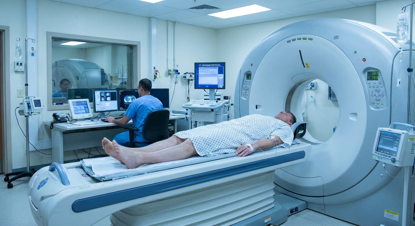
CT scan
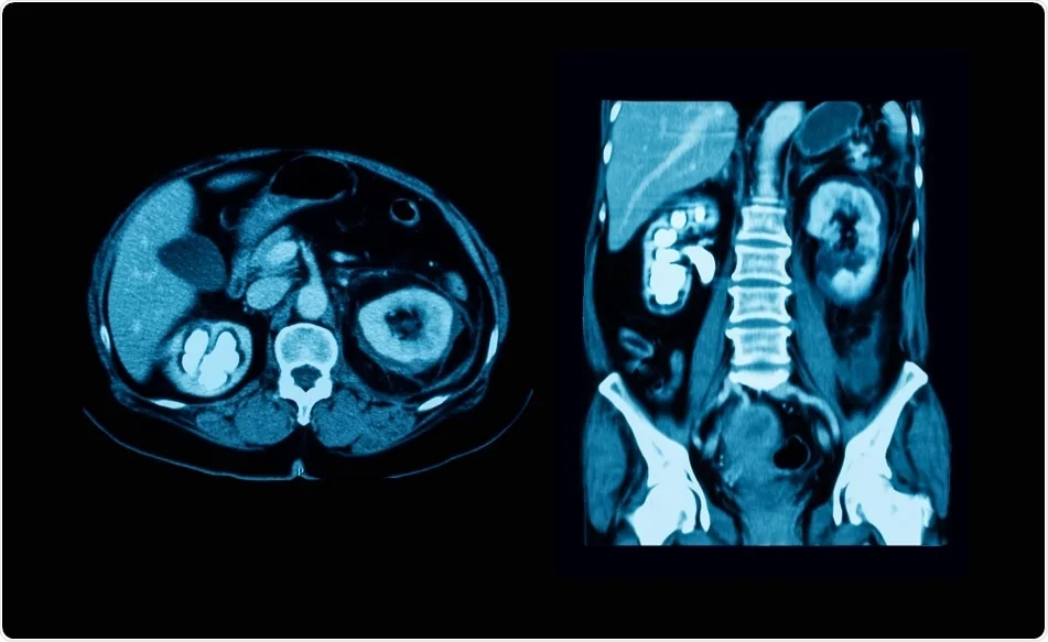
A non-contrast CT is excellent for detecting many stones. Like any test, it has limits. Very small stones can be hard to see, and some uncommon stones (for example, certain medication-related stones such as indinavir) may be radiolucent. CT can also show secondary signs of obstruction even when the stone itself is not obvious.
If a CT shows hydronephrosis but no visible stone and there is a consistent transition point where the ureter goes from dilated to narrowed, a stricture moves up the list. However, other problems can mimic this appearance, including:
- Blood clot in the collecting system or ureter
- Sloughed papilla (papillary necrosis)
- Urothelial tumor inside the ureter
- Extrinsic compression from a mass, enlarged lymph nodes, or retroperitoneal fibrosis
- Anatomic kinks or a crossing vessel pattern that can resemble a junction-level obstruction
A CT with contrast (often a CT urogram when appropriate) can help show:
- Where urine flow slows or stops
- The transition point between a dilated ureter above and a narrower segment below
- Whether there is concern for a mass, external compression, or postsurgical changes
MRI / MR urogram
MR urogram can evaluate urinary obstruction without radiation, which can be useful in select patients.
Nuclear medicine renal scan
If the question is not just “Is it narrow?” but “How much is it affecting kidney function and drainage?” a renal scan can measure how well each kidney functions and how well urine drains over time. This can be especially helpful when hydronephrosis is present but the degree of true obstruction is uncertain.
Retrograde pyelogram
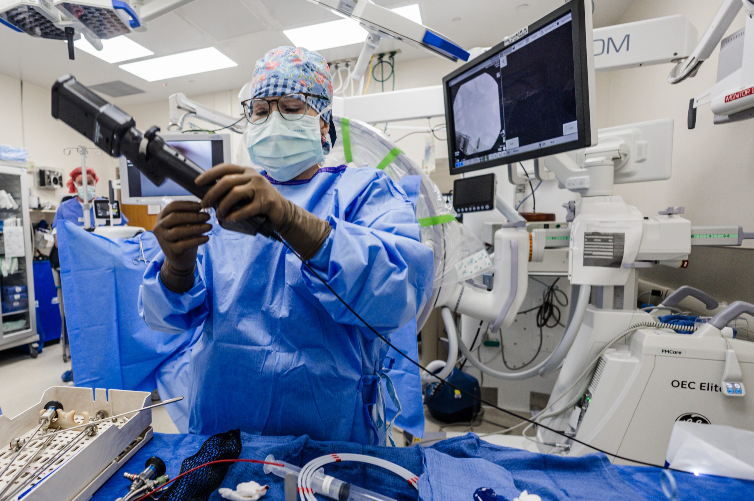
This is done by urology, usually during cystoscopy. Dye is introduced into the ureter from the bladder side. It can define the stricture location and length very clearly and is often used when planning treatment.
Stricture vs UPJ vs stone
These conditions can all cause hydronephrosis, but they are not the same problem, and the best treatment can differ.
Ureteral stricture
- Cause: scarring after stone, procedure, radiation, surgery, ischemia, or inflammation
- Pattern: a consistent bottleneck at a particular ureter segment
- Course: tends not to resolve on its own if the narrowing is significant
UPJ obstruction
Ureteropelvic junction (UPJ) obstruction is a narrowing where the kidney meets the ureter, right at the top of the drainage system. It is often congenital, though it may not be discovered until adulthood. It can also be related to an anatomic crossing vessel or, less commonly, scarring after stones or inflammation. On imaging, it typically looks like dilation of the kidney’s collecting system with less dilation further down the ureter.
A ureteral stone that may still pass
A stone can block flow, but it is not scar tissue. Many stones move over time. Symptoms often come in waves, and imaging may show a stone at a specific level with dilation above it. If the stone passes, the obstruction resolves. With a stricture, the narrowing stays put.
If imaging shows hydronephrosis but no stone, and symptoms persist or kidney function changes, it is reasonable to ask, “Could this be a stricture, or is there another cause of obstruction we need to rule out?”
Why it matters
One of the hardest parts about urinary obstruction is that it can be quiet. Some people have minimal pain but progressive swelling and declining kidney function over time.
When urine backs up for too long, it can reduce kidney function in that kidney. The goal is to relieve obstruction and protect long-term kidney health.
Treatment
Treatment depends on the stricture’s location, length, cause, severity of blockage, whether infection is present, and how well the kidney is functioning.
Urgent drainage
If there is an infection with obstruction, significant kidney injury, uncontrolled pain, or a solitary kidney, drainage may be urgent. Common options include:
- Ureteral stent: a small tube placed inside the ureter to hold it open and allow urine to drain
- Percutaneous nephrostomy tube: a tube placed through the back directly into the kidney to drain urine externally
Endoscopic treatments
- Balloon dilation: stretching the narrowed segment
- Endoureterotomy: cutting scar tissue from inside the ureter (methods vary), usually followed by a stent while it heals
These approaches can work well for short strictures in carefully selected cases. Success rates vary by stricture length, location, and cause, and recurrence is more common with longer strictures, ischemic strictures, and radiation-related scarring.
Surgical repair
For longer, recurrent, or complex strictures, urologists may recommend reconstructive surgery. The exact approach depends on where the stricture is:
- Ureteroureterostomy: removing the scarred segment and reconnecting healthy ends
- Ureteral reimplantation: reattaching the ureter into the bladder, sometimes with techniques to gain length
- More complex reconstruction when strictures are extensive
Observation
Sometimes a mild narrowing with good drainage and stable kidney function is monitored with repeat imaging and labs. This decision is individualized.
Follow-up
Follow-up is usually part of the plan, even after successful treatment. Depending on your situation, your clinician may recommend repeat labs, ultrasound or CT-based imaging, or functional testing to confirm drainage and to watch for recurrence.

What to ask
If a scan mentions hydronephrosis, narrowing, or possible stricture, these questions can help you get clarity quickly:
- Where exactly is the narrowing, and how long is it?
- Do you think this is a fixed stricture, a recently passed stone, or another cause of blockage?
- How is my kidney function right now (creatinine, eGFR), and how does it compare to my baseline?
- Is the other kidney healthy enough to compensate?
- Do I need a urology referral, and how soon?
- What symptoms should send me to the ER?
When to seek urgent care
Obstruction can become an emergency in certain situations. Get urgent evaluation if you have:
- Fever (especially with chills) plus flank pain or urinary symptoms
- Severe, uncontrolled pain or persistent vomiting
- Difficulty urinating or very low urine output
- Known kidney disease, a kidney transplant, or only one functioning kidney and new flank pain
- Worsening weakness, confusion, or signs of dehydration
An infected, obstructed kidney is particularly dangerous and needs rapid treatment.
FAQ
Can a ureteral stricture go away on its own?
True scar strictures generally do not resolve completely without intervention. Temporary swelling after a stone or a procedure can mimic a stricture early on, which is why follow-up imaging sometimes matters.
Does a stricture always cause pain?
No. Some people have minimal pain but still develop hydronephrosis and gradual loss of kidney function in that kidney.
If my creatinine is normal, does that mean I am fine?
Not necessarily. A normal creatinine can happen if the other kidney compensates. Your clinician may look at imaging severity, compare your creatinine to your personal baseline, and consider functional testing if needed.
Is a ureteral stricture cancer?
Most strictures are from scarring and are not cancer. That said, clinicians take obstruction seriously and may order imaging or procedures to be confident there is not a tumor inside or outside the ureter causing blockage.
The bottom line
A ureteral stricture is a fixed, scar-related narrowing of the ureter that can slow or block urine flow, leading to hydronephrosis, flank pain, recurrent infections, and sometimes a rise in creatinine when the other kidney cannot compensate. It is different from UPJ obstruction and different from a stone that is still moving and may pass.
If your imaging report mentions hydronephrosis without a clear stone, or you have persistent symptoms after a stone or procedure, it is worth asking directly whether a stricture is on the table, what else needs to be ruled out, and what the next step should be to protect your kidney.