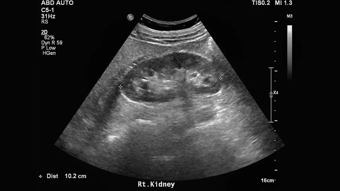
If you just read the words “extrarenal pelvis” on an ultrasound or CT report, it is completely understandable to feel uneasy, especially if the report also mentions “prominent” or “dilated” collecting system. In clinic, I often remind patients that imaging reports describe shapes and patterns, not just diagnoses. An extrarenal pelvis is usually one of those shape-based findings that sounds scarier than it is.
In most cases, an extrarenal pelvis is simply an anatomic variant, meaning it is a normal way your body is built. When imaging shows no signs of obstruction, an extrarenal pelvis by itself does not mean the kidney is blocked and typically does not require drainage.
One quick translation that can help: when a report mentions collecting system dilation, it is talking about the urine-drainage spaces inside the kidney looking more open or “full” than expected. The reason matters, and it is not always blockage.

Quick anatomy
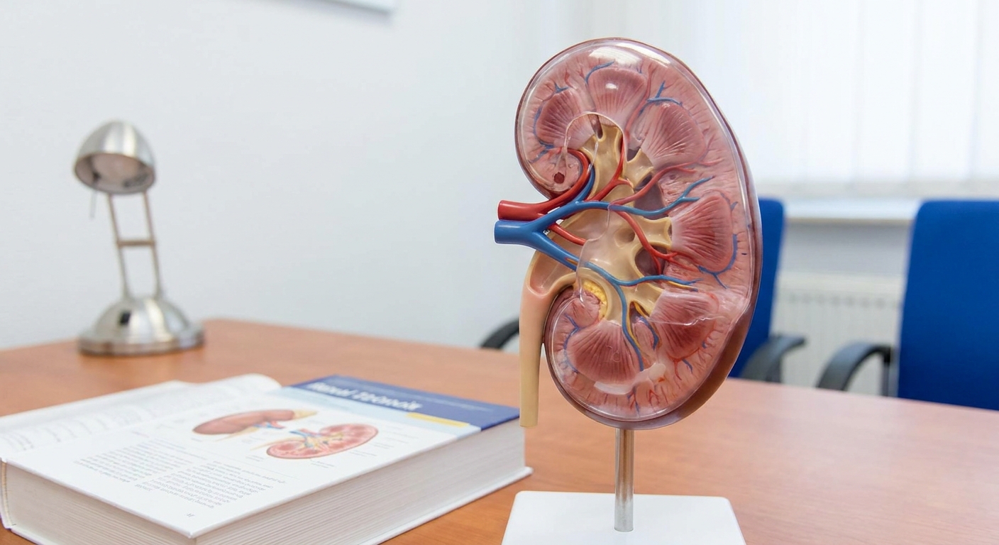
Your kidneys make urine. Inside each kidney is a branching drainage area called the collecting system. The small cups that first catch urine are the calyces (singular: calyx). These drain into a larger funnel-like space called the renal pelvis, which narrows into the ureter (the tube that carries urine to the bladder).
Most of the renal pelvis sits within the central part of the kidney, called the renal sinus. But not everyone’s anatomy is arranged the same way.
What an extrarenal pelvis is
An extrarenal pelvis means the renal pelvis sits partly outside the kidney instead of being tucked fully inside the renal sinus.
Because this pelvis is positioned more externally, it can look:
- Roomier than expected
- More visible on ultrasound
- Mildly “full” or “dilated” even when urine is flowing normally
This is why it can be mistaken for early hydronephrosis on ultrasound, especially if the report is brief or if the scan was limited by hydration status, body position, bowel gas, or a very full bladder.
It can also be bilateral (seen on both sides) and still be a benign variant.
Extrarenal pelvis vs hydronephrosis
Hydronephrosis is not just a roomy pelvis. It means urine is backing up because something is slowing or blocking drainage, such as a kidney stone, a stricture, prostate enlargement, or pregnancy-related physiologic dilation or compression.
How they look different
Radiology teams look for a pattern.
- Extrarenal pelvis: The renal pelvis may look prominent, but the calyces are not stretched (no calyceal blunting), the kidney tissue (parenchyma) is usually preserved, and there may be no ureteral dilation.
- True hydronephrosis: The dilation typically involves the calyces as well as the pelvis. Depending on severity and duration, there may be thinning of kidney tissue, and sometimes the ureter is dilated too (hydroureter) if the blockage is lower down.
One of the simplest ways to think about it is this: hydronephrosis changes the “branches” (calyces), not just the “funnel” (pelvis).
A useful nuance: very early or partial obstruction can be subtle on imaging, and sometimes the right answer comes from correlating the scan with symptoms, urine tests, and kidney function labs, or repeating imaging under better conditions.
Why it matters
Hydronephrosis can threaten kidney function if it is significant or persistent. An extrarenal pelvis, when it is purely an anatomic variant and obstruction has been excluded, usually does not require a stent, nephrostomy tube, or emergency drainage because there is no blocked urine flow to fix.
Extrarenal pelvis vs parapelvic cyst
Another common look-alike on ultrasound is a parapelvic cyst (sometimes called a renal sinus cyst). This is a fluid-filled cyst that sits near the renal pelvis. Terminology varies a bit between reports, but the practical point is the same: a cyst is a separate fluid pocket that can mimic the collecting system.
The key difference
- Extrarenal pelvis: Part of the normal collecting system. It connects to the calyces and ureter because it is literally the urine drainage pathway.
- Parapelvic cyst: A separate fluid pocket that does not connect to the collecting system.
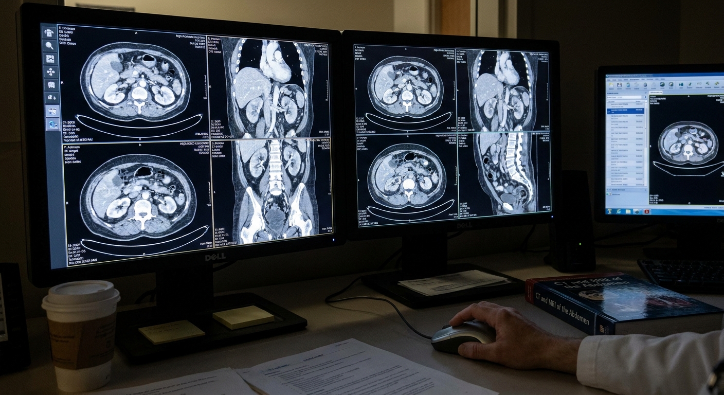
On ultrasound, both can appear as dark fluid areas in the central kidney region. And sometimes ultrasound truly cannot tell with confidence. When the picture is unclear, radiologists may recommend a follow-up study, such as a CT urogram, MR urogram, or a repeat ultrasound focused on whether there is communication with the collecting system.
Does it cause symptoms?
By itself, an extrarenal pelvis usually causes no symptoms. It is most often found incidentally when imaging is ordered for something else.
If you have symptoms like flank pain, fever, painful urination, or blood in the urine, those symptoms are more likely related to conditions such as stones, infection, or true obstruction. In other words, the symptoms deserve evaluation, but the extrarenal pelvis is often not the culprit.
Do you need treatment?
If the finding is described as a pure anatomic variant and there are no signs of obstruction, then no procedure is needed just for the anatomy.
Treatment decisions are based on whether there is evidence of obstruction or another problem, for example:
- A kidney stone blocking urine flow
- Infection plus obstruction (this is an urgent situation)
- A mass effect compressing the ureter
- Scarring or narrowing (stricture)
If your report is unclear, it is reasonable to ask your clinician: “Is there hydronephrosis, or is this just an extrarenal pelvis variant?”
Common next steps
When a scan is borderline or the report language feels vague, the next step is often something simple and practical, not a procedure.
- Review the Impression section of the report with your clinician. That is where radiologists usually state what they think it most likely is.
- Repeat ultrasound after you have emptied your bladder, or with different positioning, if a full bladder or angle may have contributed.
- Correlate with labs (creatinine, eGFR) and urinalysis, especially if there is pain, fever, or blood in the urine.
- Further imaging (CT, CT urogram, or MR urogram) if obstruction cannot be confidently excluded or if symptoms suggest a stone or blockage.
Try not to self-diagnose from one line in the Findings section. The same word, like “prominent,” can mean different things depending on the full pattern.
What to ask
You do not need to speak “radiology” to advocate for yourself. These questions are practical and usually get you a clear answer quickly:
- Do the calyces look dilated, or only the pelvis?
- Is the ureter dilated?
- Is there any sign of blockage, like a stone?
- Is kidney function normal on my labs (creatinine, eGFR)?
- Do I need repeat imaging, and if so, when and what type?
When to seek urgent care
Even if an extrarenal pelvis is usually benign, you should get urgent evaluation if you have symptoms that could signal infection or an obstructed kidney:
- Fever or chills, especially with flank or back pain
- Severe one-sided flank pain with nausea or vomiting
- Inability to urinate
- Feeling very ill or confused
- Known pregnancy with significant flank pain or fever
An infected, obstructed kidney is a medical emergency. Imaging wording should never outweigh how sick someone feels.
Common report phrases
“Prominent renal pelvis”
This can be used for either a mild anatomic fullness or early obstruction. The details of the calyces and ureter, plus your symptoms and labs, are what clarify it.
“Extrarenal pelvis noted”
This often signals the radiologist believes it is a variant, not hydronephrosis, especially if the report also states there is no calyceal dilation.
“Mild pelviectasis”
Pelviectasis means dilation of the pelvis. Sometimes it is used loosely and can overlap with an extrarenal pelvis description. If the calyces are normal, it may not be hydronephrosis.
FAQ
Is an extrarenal pelvis congenital?
Yes, it is generally considered a normal variation in how the kidney is formed. Many people have it their whole life and never know until imaging is done.
Can it turn into hydronephrosis?
The anatomy itself does not “turn into” obstruction. But someone with an extrarenal pelvis can still develop hydronephrosis later for the same reasons anyone can, such as a stone. The key is whether imaging shows calyceal dilation and whether symptoms or labs suggest blockage.
Does it affect kidney function?
Not when it is purely an anatomic variant and obstruction has been excluded. Kidney function concerns come from obstruction, repeated infections, reflux, or other kidney diseases, not from the pelvis being positioned more externally.
Should I see a urologist?
Sometimes, but not always. Referral is more likely if you have recurrent urinary infections, kidney stones, persistent flank pain, declining kidney function, or imaging that cannot confidently rule out obstruction.
Takeaway
An extrarenal pelvis is an anatomic variant where the renal pelvis sits more outside the kidney. On ultrasound it can look prominent, even when the calyces are not stretched and the ureter is not dilated. That is why it can mimic hydronephrosis.
The reassuring headline for most people is this: when it is truly just an extrarenal pelvis with no evidence of obstruction, it does not need drainage. If you are unsure what your report implies, ask whether there are signs of true obstruction, and let your symptoms and kidney function tests guide the next steps.