In clinic, I have watched the mood in the room change the moment a blockage is relieved. A patient who has been uncomfortable, nauseated, or scared suddenly breathes easier. Then the next question comes quickly and honestly: Will my kidney recover?
The answer is often encouraging, but it is not one-size-fits-all. Kidneys can rebound after an obstruction is opened, especially when it is addressed promptly. When the blockage has been there longer, or imaging shows more structural change, recovery can be partial, slower, or sometimes limited.
What happens during a blockage
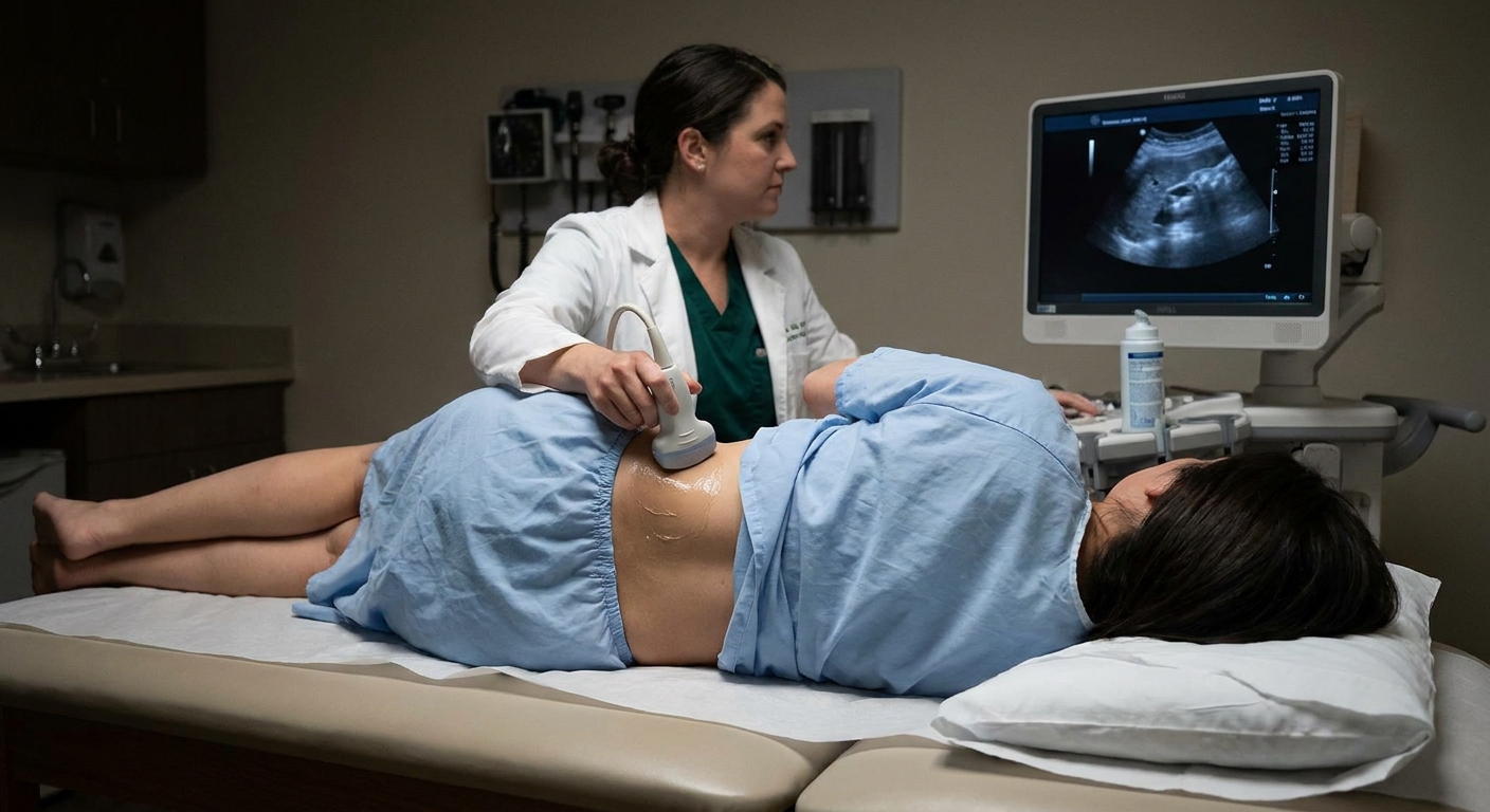
A urinary blockage can occur anywhere along the drainage path, from the kidney down through the ureter to the bladder and urethra. Common causes include kidney stones, enlarged prostate, strictures or scarring, tumors, pregnancy-related compression, blood clots, or congenital narrowing.
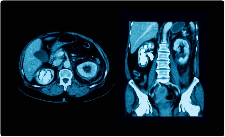
When urine cannot drain normally, pressure builds up behind the obstruction. That pressure can stretch the collecting system and cause hydronephrosis, which is the swelling you might see on ultrasound or CT. Over time, higher pressure can reduce blood flow in the kidney and strain the delicate filtering units (nephrons). The longer that pressure remains, the more likely function is to suffer.
Can function return after it is fixed?
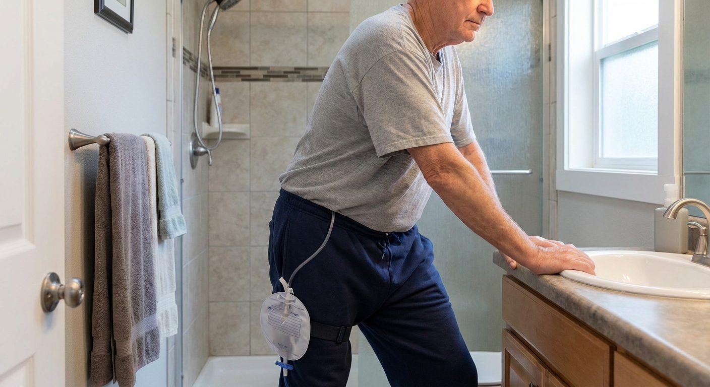
Often, yes. Many people see meaningful improvement after the blockage is relieved with a ureteral stent, nephrostomy tube, catheter, or a procedure that removes the cause (like stone removal or prostate treatment).
But kidney recovery depends on a few big factors:
- How long the blockage lasted
- Whether one kidney or both kidneys were affected
- Your baseline kidney health (age, diabetes, hypertension, prior chronic kidney disease)
- Whether infection or sepsis was present
- What the kidney looks like on imaging, especially cortical thickness
It also matters whether the obstruction was complete or partial, and whether it was an upper-tract problem (like a ureteral stone) versus bladder outlet obstruction (like an enlarged prostate). The mechanics and follow-up can differ.
Timeline after drainage
These timelines are common patterns, not promises. Recovery can be faster or slower depending on the cause, whether drainage is complete, and what your kidneys were like before the obstruction.
First 24 to 72 hours
Once the blockage is relieved, the kidney often begins to drain right away. Some people urinate a lot more than usual for a short period. This is called post-obstructive diuresis. It can be a normal response as the body clears extra salt and water.
Clinically significant post-obstructive diuresis is more likely when both kidneys were obstructed or you have a solitary functioning kidney. In those settings, monitoring may include urine output, blood pressure, and electrolytes, sometimes in the hospital, because dehydration and electrolyte shifts can happen.
Lab work, especially serum creatinine, may start improving quickly if the obstruction was the main issue and the kidney tissue is still viable.
Weeks 1 to 4
This is a common window for noticeable change on blood tests, particularly when obstruction was acute and fully relieved. Many patients see creatinine trend down during this time as inflammation settles and filtration improves. Symptoms like flank pressure, nausea, or appetite issues often improve too, although discomfort from a stent can create its own set of symptoms.
Months 1 to 6
Kidney recovery can continue for months. Some people have a quick early improvement and then slower gains. Others improve gradually the whole time. Follow-up imaging and repeated labs help clarify whether the kidney is stabilizing, continuing to recover, or showing signs of lasting damage.
If there is significant long-standing obstruction, the kidney may not return to its prior baseline. In those cases, the goal becomes protecting the function that remains and preventing another obstruction.
Why blockage duration matters
I explain this to patients using a simple analogy: if you step on a garden hose briefly, the flow bounces back. If the hose is kinked for days and the material is damaged, the flow may never be quite the same.
In the body, a shorter obstruction is more likely to be reversible. A longer obstruction is more likely to cause scarring and loss of functioning nephrons. Unfortunately, in adults, the kidney cannot simply regenerate brand-new nephrons once they are lost.
In real life, it is not always clear exactly when a blockage started. Some obstructions are painful and obvious, like a ureteral stone. Others are quiet, like slow prostate-related obstruction or a partial narrowing, and are discovered later on imaging.
Cortical thinning and recovery
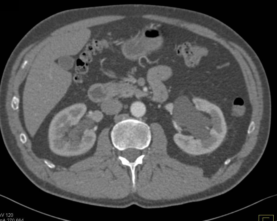
The renal cortex is the outer portion of the kidney where much of the filtering work happens. When imaging reports mention cortical thinning (or “thinned renal parenchyma”), it can be a clue that the kidney has been under stress for a while.
Generally speaking:
- Preserved cortical thickness often suggests a better chance of functional recovery after drainage.
- Marked cortical thinning raises concern for chronic injury and a lower ceiling for recovery.
Cortical thickness is a helpful proxy, but it is not perfectly specific. Thinning can also reflect other chronic processes like reflux nephropathy, ischemic disease, or other forms of medical kidney disease. That is why I interpret it in the context of your history, your labs, and how the kidney drains after treatment.
What creatinine can tell you
Creatinine is a blood test used to estimate kidney filtration. When kidneys filter less effectively, creatinine tends to rise. After a blockage is relieved, clinicians often follow creatinine closely because it is one of the quickest objective indicators of recovery.
One important nuance is that creatinine and eGFR are influenced by muscle mass and overall body size. That is one reason clinicians focus on trends rather than a single value.
Helpful patterns
- Creatinine drops after drainage: suggests the obstruction was significantly affecting kidney filtration and that at least some function is returning.
- Creatinine plateaus above your prior baseline: may mean partial recovery, underlying chronic kidney disease, or persistent issues (like incomplete relief or additional kidney stressors).
- Creatinine stays high or worsens: can indicate ongoing obstruction, infection, dehydration, medication effects, or intrinsic kidney injury that needs urgent evaluation.
Limits to know
Creatinine reflects how both kidneys are doing together, not just the obstructed one. If only one kidney was blocked and the other kidney is healthy, creatinine may be normal even if the obstructed kidney is struggling.
A practical example: a single ureteral stone blocking the left kidney can hurt badly, but creatinine may stay near normal if the right kidney is healthy. In contrast, bladder outlet obstruction (like an enlarged prostate) can affect both kidneys and is more likely to push creatinine up.
Follow-up scans after relief
Follow-up imaging is not only about confirming improvement in swelling. It is also about ensuring the kidney is draining, checking for persistent dilation, and looking for structural changes that guide next steps.
Also, pain relief does not always equal full decompression. A stent can migrate, kink, or become obstructed. A stone can be residual. That is another reason follow-up matters even when you feel better.
Common follow-up tests
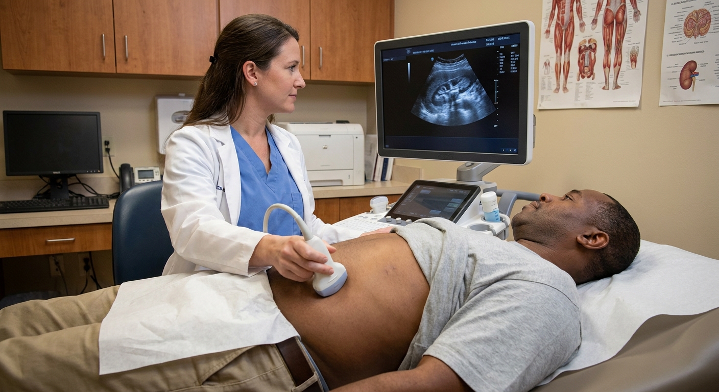
- Renal ultrasound: Often used because it is noninvasive and avoids radiation. It can show persistent hydronephrosis, improvement, or worsening.
- CT (in selected cases): Helps identify stones, masses, or anatomic concerns when ultrasound is unclear.
- Nuclear medicine renal scan (often called a MAG3 or DTPA scan): Can estimate how much each kidney contributes to overall function and whether drainage is delayed. This can be very helpful when deciding whether an obstructed kidney is recovering or has limited remaining function.
The timing depends on the situation. Some people need imaging within weeks, while others are followed over a few months, particularly when swelling takes time to resolve even after the obstruction is fixed.
What partial damage can look like
Many patients assume recovery is either complete or not possible. In reality, partial recovery is common.
Partial recovery may mean:
- You regain enough kidney function to return to a stable creatinine, but not quite your old baseline.
- The kidney drains better, swelling improves, and pain resolves, but imaging shows residual thinning.
- One kidney remains weaker, but the other kidney compensates well, so day-to-day life is normal.
If a kidney has limited recovery, the focus shifts to protecting the function you have: controlling blood pressure, managing diabetes carefully, avoiding dehydration, and reviewing medications that can strain kidneys (like NSAIDs such as ibuprofen or naproxen, when alternatives are appropriate).
Red flags after drainage
After an obstruction is relieved, complications are still possible. Contact your clinician urgently or seek emergency care if you have:
- Fever, chills, or confusion (possible infection)
- Worsening flank pain that is severe or persistent
- Vomiting or inability to keep fluids down
- Little to no urine output, especially if you were told both kidneys were affected
- Blood in urine that is heavy, worsening, or associated with clots and urinary retention
- Dizziness, fainting, or extreme thirst after very high urine output (possible dehydration from post-obstructive diuresis)
Questions to ask at follow-up
- What caused the obstruction, and how do we prevent it from happening again?
- Was one kidney affected or both?
- Did imaging show cortical thinning or scarring?
- How often should I recheck creatinine and electrolytes?
- Do I need follow-up ultrasound or a nuclear medicine renal scan?
- If I have a stent or nephrostomy tube, what symptoms are expected and what symptoms are not?
FAQ
How long does it take for creatinine to improve after a blockage is relieved?
It varies. Improvement may begin within days and continue over several weeks. Some recovery can continue for months, especially if the obstruction was significant but treated in time and drainage is complete.
If my ultrasound still shows hydronephrosis, does that mean I am still blocked?
Not always. Swelling can take time to settle even after drainage is restored, and some people have residual dilation without active obstruction. That is why your clinician pairs imaging with symptoms, creatinine trends, and sometimes functional tests.
Can a kidney fully recover after hydronephrosis?
Sometimes, yes, especially when the obstruction is short-lived and promptly relieved. If the obstruction was long-standing or imaging shows marked cortical thinning, recovery is more likely to be partial.
If only one kidney was blocked, could my bloodwork still look normal?
Yes. A healthy opposite kidney can compensate, so creatinine may stay in the normal range even if the obstructed kidney has reduced function. Your care team may rely more on imaging and kidney-specific functional testing in that situation.
The bottom line
A kidney can often recover after a blockage is relieved, and the most meaningful improvements commonly happen in the first few weeks, with possible continued gains over months. The biggest predictors are how long the obstruction lasted, whether drainage is complete, and whether imaging suggests chronic change, such as cortical thinning. Trends in creatinine and the right follow-up scans help your clinician estimate recovery and plan next steps.
If you are walking through this right now, please know this: you are not behind for asking questions. Understanding what your labs and imaging mean is part of protecting your kidneys for the long run.