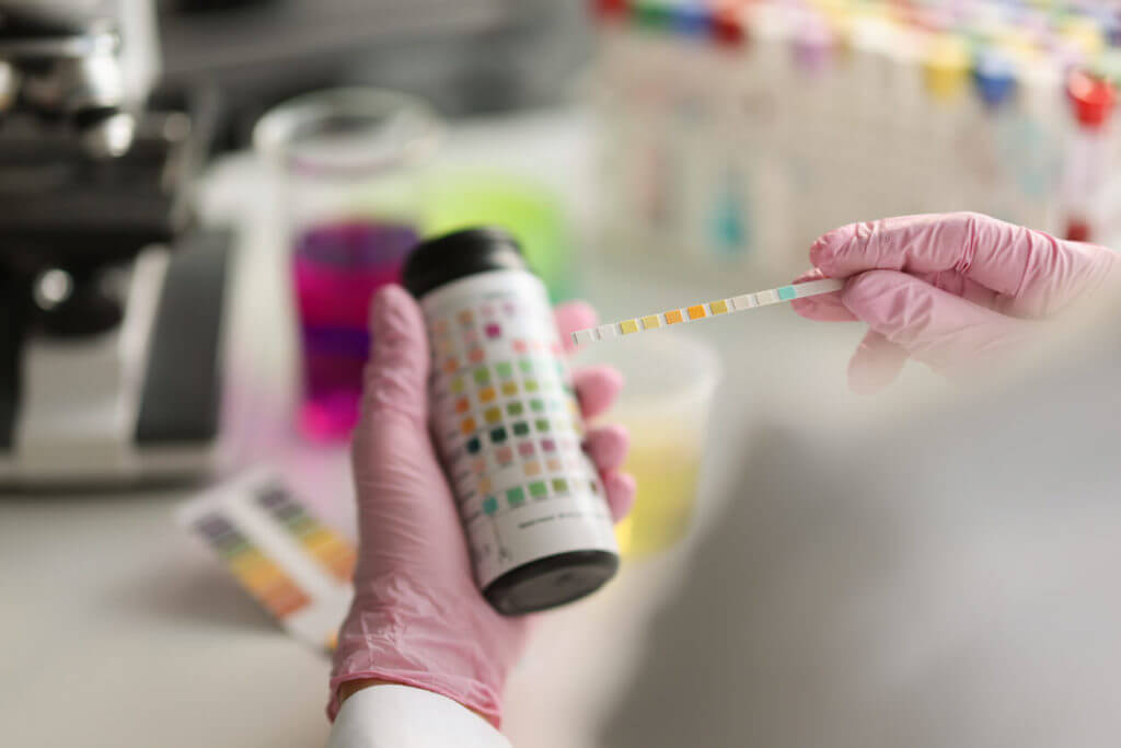
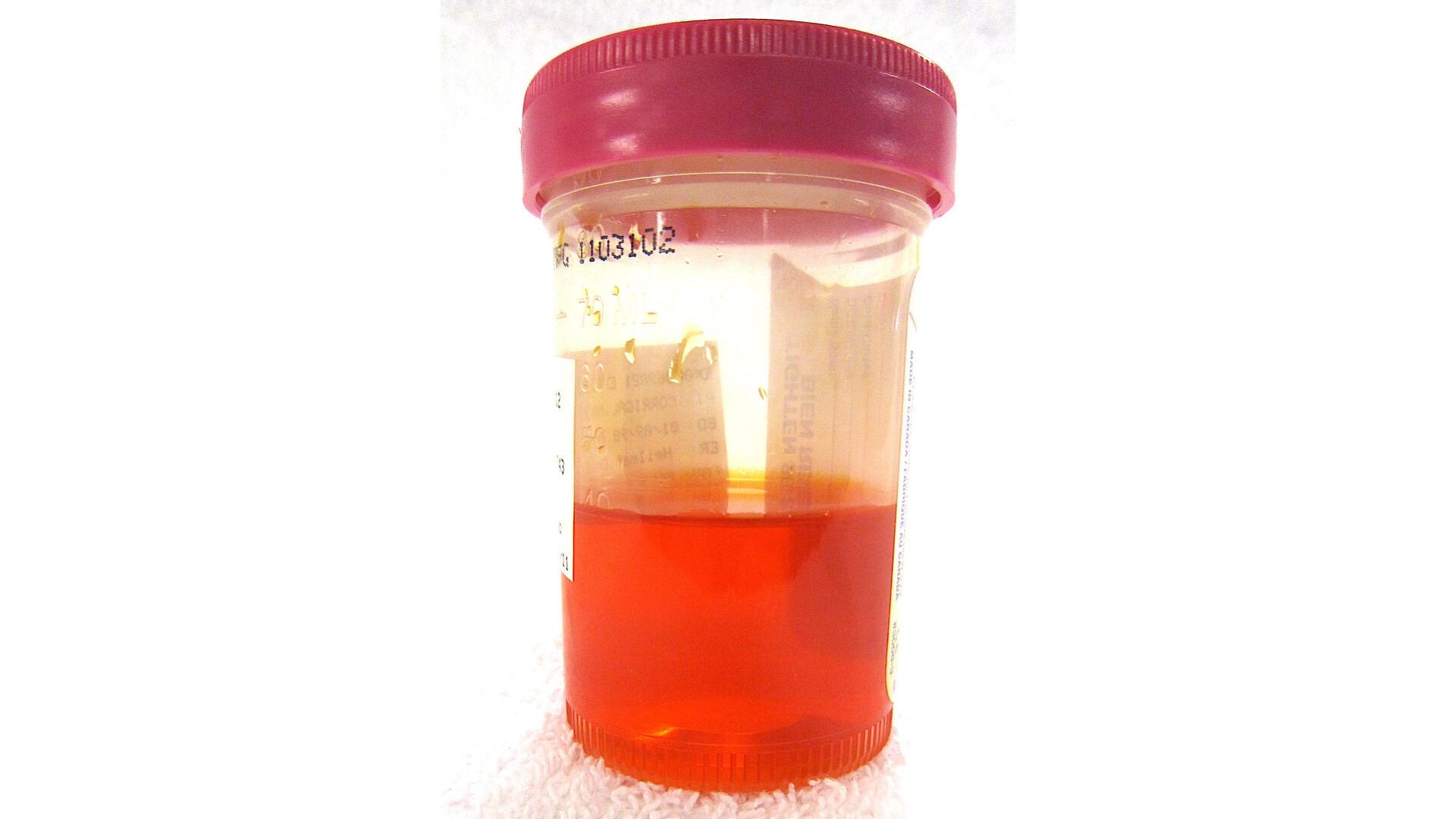
In my family clinic days, one of the most common sources of anxiety I saw was a lab portal notification that simply said: Urinalysis: abnormal. It is understandable. A urinalysis report can look like a wall of unfamiliar terms, and yet it is one of the most useful, everyday tests we have for spotting dehydration, urinary tract infections, kidney issues, diabetes clues, and sometimes causes of blood in the urine.
One reassuring truth up front: an “abnormal” flag is common, and many abnormalities are minor, temporary, or related to how the sample was collected rather than a serious problem.
A urinalysis (often shortened to UA) is a set of checks done on a urine sample. Most UAs include three parts:
- Visual exam (what it looks like)
- Chemical dipstick (a strip that changes color based on what is in the urine)
- Microscopic exam (looking under the microscope for cells, crystals, bacteria, or casts)
This article walks through common urinalysis lines one by one, explains what “normal” tends to look like, and clarifies when results need follow-up.

Before you read the numbers
Collection matters
Many confusing results come down to collection. The usual outpatient method is a clean-catch midstream sample. That helps reduce contamination from skin cells, vaginal discharge, semen, or bacteria at the urethral opening.
- Menstruation can cause blood and red blood cells to show up even when the urinary tract is fine.
- Recent sex can sometimes irritate tissue and affect blood or white cells.
- Heavy exercise can transiently cause blood or protein in urine.
- Delay in testing can change results. For example, bacteria can multiply and urine pH can rise if a sample sits too long at room temperature. Cells and casts can also break down, and some dipstick readings may be less reliable.
Dipstick vs microscopy
Dipstick results are fast and helpful, but they are not perfect. When something important is positive on the dipstick, clinicians often confirm it with the microscopic portion (or repeat the test) before making big conclusions.
How to read a urinalysis report
Different labs format reports differently, but most include the same core items.
Color
What it is: A quick visual check.
Common “normal”: Pale yellow to amber.
What changes can mean:
- Very dark yellow often points to dehydration (more concentrated urine).
- Red or pink can be blood, but can also be foods (beets, blackberries) or medications (phenazopyridine, rifampin).
- Brown or tea-colored may be dehydration, liver issues with bilirubin, or muscle breakdown (myoglobin). This needs medical attention if new.
- Cloudy can be infection, crystals, mucus, semen, or contamination.
Specific gravity
What it is: How concentrated the urine is. It reflects hydration status and, indirectly, how well the kidneys are concentrating urine.
Typical range: About 1.005 to 1.030 (ranges vary by lab).
How to think about it:
- Low specific gravity can occur with very high fluid intake or conditions that reduce the kidney’s ability to concentrate urine.
- High specific gravity often suggests dehydration. It can also rise with glucose or protein in urine.
pH
What it is: How acidic or alkaline the urine is.
Typical range: Around 4.5 to 8.0.
What shifts can mean:
- More acidic urine can show up with high-protein diets, dehydration, diarrhea, or certain metabolic states.
- More alkaline urine can show up with some UTIs (certain bacteria raise pH), vomiting, or it can be higher after meals in some people.
Urine pH can matter for kidney stone risk because some stones form more easily at certain pH levels.
Blood (heme) on dipstick
What it is: The dipstick “blood” pad reacts to the heme component found in hemoglobin (from red blood cells) and in myoglobin (from muscle).
Important nuance: A positive dipstick for blood does not always mean you are truly peeing out intact red blood cells.
Dipstick blood with no RBCs
If the dipstick is positive for blood, but the microscope shows few or no red blood cells, clinicians consider a few possibilities:
- Hemoglobinuria: free hemoglobin in urine, which can happen with hemolysis (breakdown of red blood cells in the bloodstream).
- Myoglobinuria: myoglobin from muscle injury or severe muscle breakdown (rhabdomyolysis). This can be serious, especially if accompanied by muscle pain, weakness, or dark cola-colored urine.
- False positives: oxidizing agents or contamination can rarely trigger the pad.
In clinic, this pattern is one reason we do not stop at dipstick alone. We look at the whole story: symptoms, color of urine, kidney function tests, medication and exercise history, and sometimes a repeat UA.
Hematuria (RBCs on microscopy)
Hematuria means red blood cells (RBCs) are actually present in the urine. It can be:
- Microscopic: you cannot see it, but the microscope detects RBCs.
- Gross: visible pink, red, or brown urine.
Many clinicians define microscopic hematuria as about 3 or more RBCs per high power field (HPF) on a properly collected specimen, especially if it persists.
Causes range from very treatable (UTI, kidney stones) to conditions that need prompt evaluation (certain kidney diseases, urinary tract tumors, significant trauma). The level of concern depends on your age, risk factors, symptoms, and whether hematuria persists on repeat testing.
Protein
What it is: The dipstick primarily detects albumin, a key blood protein.
How it is often reported: Many labs use grades like negative, trace, 1+, 2+, 3+ (the exact cutoffs vary by lab and by urine concentration).
What small amounts can mean: Temporary protein can show up with fever, dehydration, stress, or heavy exercise.
When it matters more: Persistent protein, especially moderate to large amounts, can be a sign of kidney disease. Clinicians often follow up with a urine albumin-to-creatinine ratio (ACR) or protein-to-creatinine ratio, because those are better ways to quantify protein loss.
Leukocyte esterase
What it is: A marker associated with white blood cells (WBCs).
What it suggests: Inflammation or infection in the urinary tract.
Common caveats: It can be positive with contamination from vaginal secretions, or with noninfectious inflammation. It can also be falsely negative if urine is very dilute or the sample is handled poorly. That is why symptoms and the microscopic WBC count matter.
Nitrites
What it is: Some bacteria convert nitrates (normally present from diet) into nitrites. A positive nitrite test supports bacterial presence.
How to interpret it:
- Positive nitrites can point toward a UTI, especially when paired with symptoms and leukocyte esterase.
- Negative nitrites do not rule out UTI. Not all bacteria make nitrites, and urine needs to sit in the bladder long enough for conversion to occur. Very frequent urination and some supplements or high vitamin C intake can also contribute to nitrite-negative results in some situations.
Glucose
What it is: Sugar in the urine.
What it usually means: Blood glucose is high enough that the kidneys spill glucose into urine, often seen with diabetes or significant hyperglycemia. It can also appear in pregnancy or with rare kidney tubule conditions.
If glucose is present, your clinician typically checks a blood glucose and often an A1C.
Other dipstick lines
- Ketones: can appear with fasting, vomiting, low-carbohydrate diets, or uncontrolled diabetes (especially type 1). If you have significant ketones and feel ill, seek urgent evaluation.
- Bilirubin and urobilinogen: can suggest liver or bile duct issues in the right context.
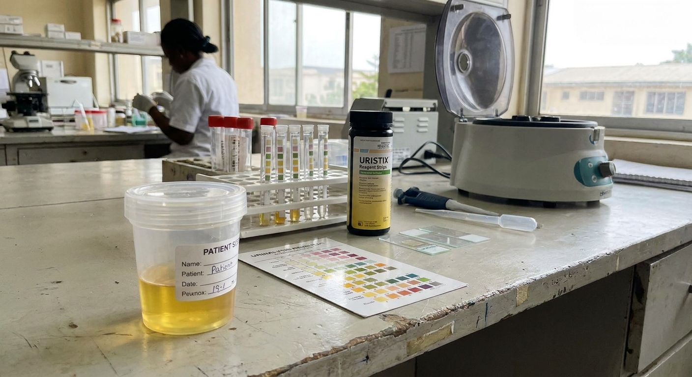
Microscopy results
If your urinalysis includes microscopy, you will often see results listed as “per high power field” (HPF) or “per low power field” (LPF). These are just microscope magnifications. As a practical example, you might see RBCs reported as “RBC 0 to 2/HPF” and casts reported as “hyaline casts 0 to 2/LPF.”
Important: “Normal” ranges vary by lab and by the situation, so use the reference ranges on your report when you have them. Still, a few rough guideposts help people make sense of common wording.
Typical ranges you may see
- RBCs: often 0 to 2/HPF is considered typical. Persistent results around 3 or more/HPF often trigger a closer look.
- WBCs: often 0 to 5/HPF is considered typical. Higher counts can support infection or inflammation in the right context.
- Squamous epithelial cells: “few” can happen even with good collection, but “many” or “moderate” often suggests contamination, meaning other lines of the UA may be less reliable.
Red blood cells (RBCs)
What they indicate: True hematuria when elevated. The pattern can help with next steps.
- RBCs plus protein and casts can raise concern for a kidney (glomerular) source.
- RBCs without much else might fit with stones, infection, trauma, prostate issues, or other urinary tract causes.
White blood cells (WBCs)
What they indicate: Inflammation. When paired with symptoms, bacteria, and positive leukocyte esterase, WBCs support UTI. WBCs can also show up with kidney stones, interstitial cystitis, sexually transmitted infections affecting the urinary tract, or contamination.
Epithelial cells
What they indicate: Often contamination, especially when “squamous epithelial cells” are present in higher amounts. In other words, the sample may have picked up skin cells, and some other UA lines are less reliable.
Bacteria and yeast
Bacteria: Can support UTI when symptoms are present, but bacteria can also show up from contamination. A urine culture is what identifies the specific organism.
Yeast: May be contamination, or it may suggest a yeast infection or yeast in urine, especially in people with diabetes or urinary catheters. Clinicians interpret this carefully and do not automatically treat it without the right context.
Crystals
Crystals can show up in people without symptoms. Certain crystal types and repeated findings may help explain kidney stones or metabolic issues. If you have flank pain or repeated stones, your clinician may order additional urine testing beyond a basic UA.
Casts
What they are: Tiny tube-shaped molds formed in the kidney’s tubules. Think of them as a snapshot of what was happening inside the kidney.
- Hyaline casts: can happen with dehydration or after exercise and may be benign.
- RBC casts: can suggest bleeding from within the kidney, sometimes seen in glomerulonephritis and needs prompt evaluation.
- WBC casts: can show up with kidney infection (pyelonephritis) or inflammatory kidney conditions.
- Granular or waxy casts: can be associated with more significant kidney disease in the right clinical picture.
Cast interpretation is one place where the overall context and sometimes repeat or confirmatory testing really matters.
UA vs culture vs cytology
These tests sound similar, but they answer different questions.
Urinalysis (UA)
- Best for: A broad screen for infection clues, blood, protein, glucose, hydration status, and microscopic findings (cells, crystals, casts).
- Timing: Fast. Dipstick results can be available within minutes. Microscopy may take longer depending on the lab.
- Limits: It cannot reliably tell you which bacteria are causing infection or which antibiotic will work.
Urine culture
- Best for: Confirming a bacterial UTI, identifying the organism, and determining antibiotic susceptibility.
- Timing: Commonly 1 to 3 days for final results (some sensitivities finalize closer to 48 to 72 hours).
- When it is often ordered: Pregnancy, complicated infections, recurrent UTIs, certain male UTIs, severe symptoms, treatment failure, or higher-risk patients.
Urine cytology
- Best for: Looking for abnormal cells that can be associated with urinary tract cancers, especially higher-grade bladder cancer.
- Timing: Varies by lab.
- Important limits: Cytology does not diagnose all cancers, and a “negative” result does not always rule out disease. It is typically used as part of a bigger evaluation plan when there is persistent hematuria or other risk factors.

When to call your clinician
Some abnormal lines are common and temporary. Others should be followed up quickly, especially if you have symptoms.
Get urgent care if you have
- Visible blood in urine, especially with clots
- Fever, chills, back or flank pain, nausea or vomiting (possible kidney infection)
- Severe pain that comes in waves (possible stone)
- Dark cola-colored urine with muscle pain or weakness (possible myoglobin)
- Signs of dehydration with inability to keep fluids down
Make an appointment if you notice
- Persistent microscopic blood on repeat testing
- Protein in urine that does not resolve, especially with swelling, foamy urine, or high blood pressure
- Recurrent UTI-type symptoms, or symptoms with negative cultures
- New urinary symptoms if you are pregnant, immunocompromised, or have kidney disease
If you are unsure, bring your actual results to the visit and ask: “Which findings matter most for my symptoms and risk factors, and do we need to repeat the test or add a culture?” That single question often clarifies the next step.
Quick FAQ
Can dehydration cause abnormal UA results?
Yes. Dehydration can concentrate urine, raising specific gravity and sometimes making trace protein or hyaline casts appear. If dehydration is suspected, clinicians may recommend rechecking after good hydration.
If my UA suggests a UTI, do I always need antibiotics?
Not always. Symptoms matter. A UA can look “infected” due to contamination or noninfectious inflammation. Also, some people have asymptomatic bacteriuria, meaning bacteria show up but there are no urinary symptoms. In many nonpregnant adults, that is often not treated. If symptoms are classic and the UA supports it, treatment is common. If the story is unclear, a urine culture can help confirm infection and guide antibiotic choice.
Why would my dipstick show blood but the microscope shows no RBCs?
Because the dipstick reacts to heme from hemoglobin or myoglobin, not only intact red blood cells. That pattern can occur with hemolysis, muscle injury, or a false positive, and it deserves a clinician’s interpretation in context.
Should I repeat an abnormal urinalysis?
Often, yes. A repeat clean-catch sample can be very helpful, especially if contamination is likely (lots of epithelial cells), you were on your period, you were dehydrated, you exercised hard, or results do not fit your symptoms.