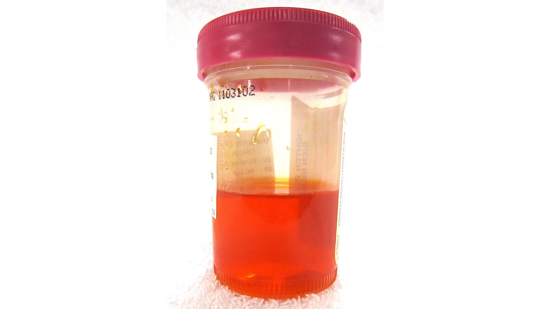
When someone tells me they have blood in their urine, their mind often jumps straight to kidney stones or cancer. Those concerns deserve respect and a prompt medical workup. But there is another, less talked about cause that can be both alarming and surprisingly mechanical: nutcracker syndrome.
Nutcracker syndrome is primarily a blood flow and pressure problem, not an infection. It is also not the same thing as a classic “kidney filtering” (glomerular) disease, though real life is messy and people can have more than one issue at once. Nutcracker syndrome happens when a vein draining the left kidney gets squeezed in a tight space in the abdomen. That squeeze can raise pressure in the vein, irritate delicate vessels, and lead to symptoms like pain and blood in the urine.
Nutcracker syndrome, explained simply
Your kidneys filter blood and make urine. To keep working, they need a steady in-and-out flow of blood.
The left renal vein is the main “exit lane” that carries blood away from the left kidney back toward the heart. In many people, that vein passes between two large arteries:
- The aorta (the body’s main artery)
- The superior mesenteric artery (SMA) (an artery that branches off the aorta and supplies part of the intestines)
In classic (sometimes called anterior) nutcracker syndrome, the left renal vein is compressed between the SMA and the aorta. Think of it like a garden hose caught between two heavy boards. Flow can still happen, but the pressure builds behind the pinch point.
There are also variants worth knowing about:
- Posterior nutcracker syndrome: the left renal vein is compressed between the aorta and the spine, often when the vein runs behind the aorta (a “retroaortic” vein).
- Circumaortic (collar) renal vein anatomy: the vein has more than one channel around the aorta, and one channel can be the one getting compressed.
You may also hear:
- Nutcracker phenomenon: the vein looks compressed on imaging, but the person has no symptoms.
- Nutcracker syndrome: compression plus symptoms that match.
Why it can cause blood in the urine
Hematuria means blood in the urine. When pressure rises in the left renal vein, smaller veins in and around the kidney can become stretched and fragile. Tiny vessels can rupture, leading to bleeding into the urine.
Blood in the urine can be:
- Visible (gross hematuria): urine looks pink, red, or cola-colored. (Cola-colored urine can also come from other causes, so it is a reason to get checked rather than to assume.)
- Microscopic: urine looks normal, but blood shows up on a lab test.
Some people also develop protein in the urine, sometimes more noticeable after being upright or active (often described as orthostatic proteinuria). In nutcracker syndrome, this protein leakage may be related to pressure changes in the kidney’s drainage system, but the mechanism is not perfectly settled and protein in urine always deserves a thoughtful evaluation.
Common symptoms
Symptoms vary a lot. Some people have intermittent, mild symptoms. Others have pain or bleeding that disrupts daily life.
Left flank or abdominal pain
This is often described as an ache or pressure on the left side of the back or abdomen. It may worsen with activity, standing for long periods, or certain positions.
Blood in the urine
This can come and go. If bleeding is heavier or persistent, it can contribute to iron deficiency anemia, leaving you feeling tired, weak, or short of breath with exertion.
Pelvic congestion symptoms
Because the pressure backs up into connecting veins, some people develop enlarged veins in the pelvis. Symptoms can include:
- Deep pelvic aching or heaviness
- Pain with or after sex
- Worsening pelvic discomfort after standing or at the end of the day
Varicocele, especially on the left
A varicocele is enlarged veins in the scrotum, often described as feeling like a “bag of worms.” It is more common on the left side because the left testicular vein drains into the left renal vein. When pressure is high in the left renal vein, a varicocele can enlarge or become symptomatic.

Who gets it?
Nutcracker syndrome is considered uncommon, and it can be underdiagnosed because symptoms overlap with more familiar problems. At the same time, it can also be over-suspected if imaging shows compression that is actually a symptom-free “phenomenon.”
It is often discussed in:
- Adolescents and young adults, sometimes during periods of rapid growth
- People with low body fat or recent significant weight loss, which can reduce the cushioning around vessels
- People with connective tissue differences or anatomy that creates a narrower angle between the SMA and aorta
That said, it can occur outside these groups too. Symptoms and the overall clinical picture matter more than any single risk factor.
How doctors diagnose it
Diagnosis is tricky for a simple reason: lots of people can have some left renal vein compression on imaging and feel fine. So clinicians have to connect the dots between symptoms, urine findings, and a vascular pattern on imaging, while also ruling out more common and dangerous causes of hematuria.
Step 1: Confirm hematuria and look for clues
- Urinalysis to confirm blood and check for protein, infection markers, and other findings
- Microscopy to look at red blood cell shape (this helps separate “glomerular” bleeding from non-glomerular sources)
- Blood tests as needed (kidney function, blood counts if bleeding is significant)
Step 2: Do the standard hematuria workup
Before anyone labels hematuria as nutcracker syndrome, many patients need the usual evaluation for urinary tract bleeding based on age, risk factors, and whether blood is visible. That often means urology involvement, and in many adult cases it may include cystoscopy and imaging to exclude bladder and kidney tumors, stones, and other structural causes.
Step 3: Imaging focused on the renal vein
Imaging choices vary based on symptoms and local practice, but commonly include:
- Doppler ultrasound: can estimate blood flow and look for signs of narrowing and higher velocities at the compression point.
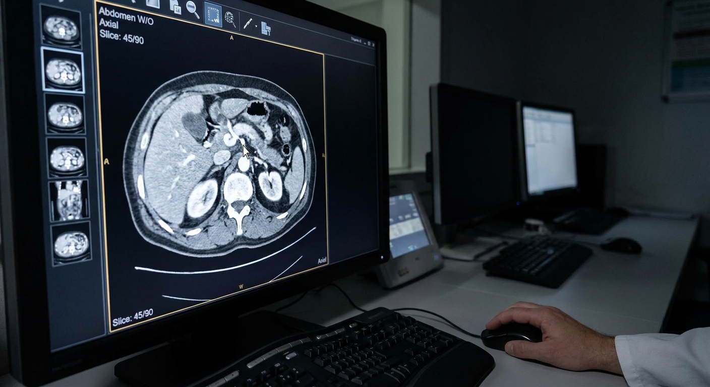
- CT angiography (CTA) or MR angiography (MRA): can show anatomy, including narrowing of the left renal vein at the compression point and enlargement upstream, plus collateral veins.
- Venography with pressure measurements (more invasive): sometimes used when the diagnosis is uncertain or when planning intervention. Many specialists consider this the closest thing to a reference standard, but pressure gradient thresholds vary, and results still need to fit the clinical picture.
The key is that nutcracker syndrome has a vascular pattern on imaging. Typically it is not explained by a stone blocking a ureter or a mass in the kidney, though incidental findings can coexist, which is another reason not to jump to conclusions from any single scan.
How it compares to other causes
Several conditions can cause blood in the urine. Here is how clinicians often separate them.
Vs. kidney stones
Kidney stones often cause sudden, severe colicky pain that can start in the flank and radiate to the groin. Nausea and vomiting are common. The pain usually comes in waves.
Nutcracker syndrome more often causes a persistent ache or pressure (though it can fluctuate) and may be associated with pelvic symptoms or a left varicocele. Imaging typically does not show an obstructing stone, but instead shows vein compression and collateral veins.
Vs. kidney or bladder tumor
Tumors in the kidney or bladder can cause hematuria, and some are silent early on. Visible blood in the urine always deserves evaluation to rule out malignancy, especially in older adults or those with risk factors like smoking.
Nutcracker syndrome is not a growth in the kidney. The imaging story centers on blood vessels and flow patterns, but the hematuria workup still needs to be completed appropriately rather than assumed.
Vs. glomerular disease
Glomerular bleeding (from diseases affecting the kidney’s microscopic filters) often shows:
- Protein in the urine (sometimes significant)
- Dysmorphic red blood cells (oddly shaped cells under the microscope)
- Red blood cell casts (tube-shaped clumps formed in the kidney tubules)
- Sometimes high blood pressure, swelling, or abnormal kidney function
Nutcracker syndrome is typically a non-glomerular cause of bleeding. Protein can occur, but the overall urine microscopy, kidney function, and vascular imaging pattern often point away from a primary filter disorder. Still, overlap is possible, and persistent protein or abnormal kidney function should never be waved away.
Treatment options
Treatment depends on symptom severity, how much bleeding is happening, and how much nutcracker syndrome is affecting quality of life. It also depends on age and anatomy, because some interventions have different risk profiles in younger patients.
Conservative management
For mild or intermittent symptoms, especially in younger patients, clinicians may recommend:
- Observation with follow-up
- Managing anemia with iron if needed
- Nutrition and weight stabilization if low body weight is a factor
- Pain management strategies individualized to the patient
Some people improve over time as body composition changes or anatomy shifts with growth.
Procedures and surgery
For more severe, persistent symptoms, recurrent significant hematuria, or complications, specialists may discuss options such as:
- Endovascular stenting of the left renal vein in select cases
- Surgical approaches such as transposition of the left renal vein or other reconstructive techniques
Stenting can be effective, but it is not a casual decision. Depending on the case, discussions may include risks like stent migration, thrombosis, need for blood thinners, and uncertainty about long-term durability, with extra caution in children and teens who are still growing.
These decisions are typically made with a vascular surgeon, interventional radiologist, and urologist or nephrologist, depending on the dominant symptoms.
If pelvic congestion or varicocele is a major issue, treatment may also involve targeted management of those dilated veins, but the underlying venous pressure problem must be considered to avoid incomplete relief.
What to expect
With any approach, follow-up matters. Symptom improvement rates vary, and some people need repeat urine testing, anemia monitoring, or follow-up imaging to make sure the plan is working and to catch other causes of hematuria that can declare themselves over time.
When to seek care urgently
Please get urgent medical attention if you have:
- Visible blood in the urine, especially with clots
- Severe flank pain, fever, or vomiting
- Symptoms of significant blood loss such as fainting, chest pain, marked shortness of breath, or extreme weakness
- Inability to urinate or worsening lower abdominal pain (possible blockage)
Even when nutcracker syndrome is the eventual explanation, the first step is always to rule out more common and potentially dangerous causes.
Common questions
Is nutcracker syndrome dangerous?
It can be very uncomfortable and scary, especially when blood appears in the urine. Many cases are not immediately dangerous, but complications like anemia, significant pain, or reduced quality of life can be serious. It also matters that hematuria can signal other conditions, so proper evaluation is essential.
Can it cause chronic pelvic pain?
Yes. Increased pressure can lead to pelvic venous congestion, which can cause heaviness or aching that worsens with prolonged standing or later in the day.
Can it cause a varicocele?
Yes, particularly on the left. A new or worsening varicocele, especially alongside hematuria or left-sided flank pain, is one reason clinicians consider nutcracker syndrome.
What specialist treats it?
Evaluation often starts with primary care or urgent care, then may involve urology (hematuria workup), nephrology (kidney and urine findings), and vascular surgery or interventional radiology (vascular diagnosis and interventions).
The bottom line
Nutcracker syndrome is a condition where the left renal vein is compressed, most commonly between the aorta and the superior mesenteric artery, but sometimes in other anatomic variants. That compression can raise venous pressure and lead to blood in the urine, left flank pain, and sometimes pelvic congestion or a left-sided varicocele.
It is also a diagnosis that should be made carefully. Imaging can show vein compression in people with no symptoms, and hematuria can come from stones, tumors, infection, or glomerular disease. If you notice blood in your urine, do not try to self-diagnose. Get checked. With the right evaluation, you can move from fear and uncertainty to a clear plan.