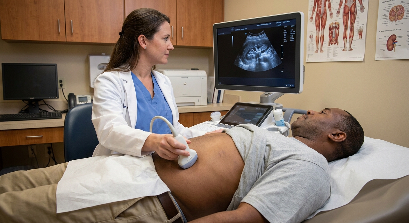
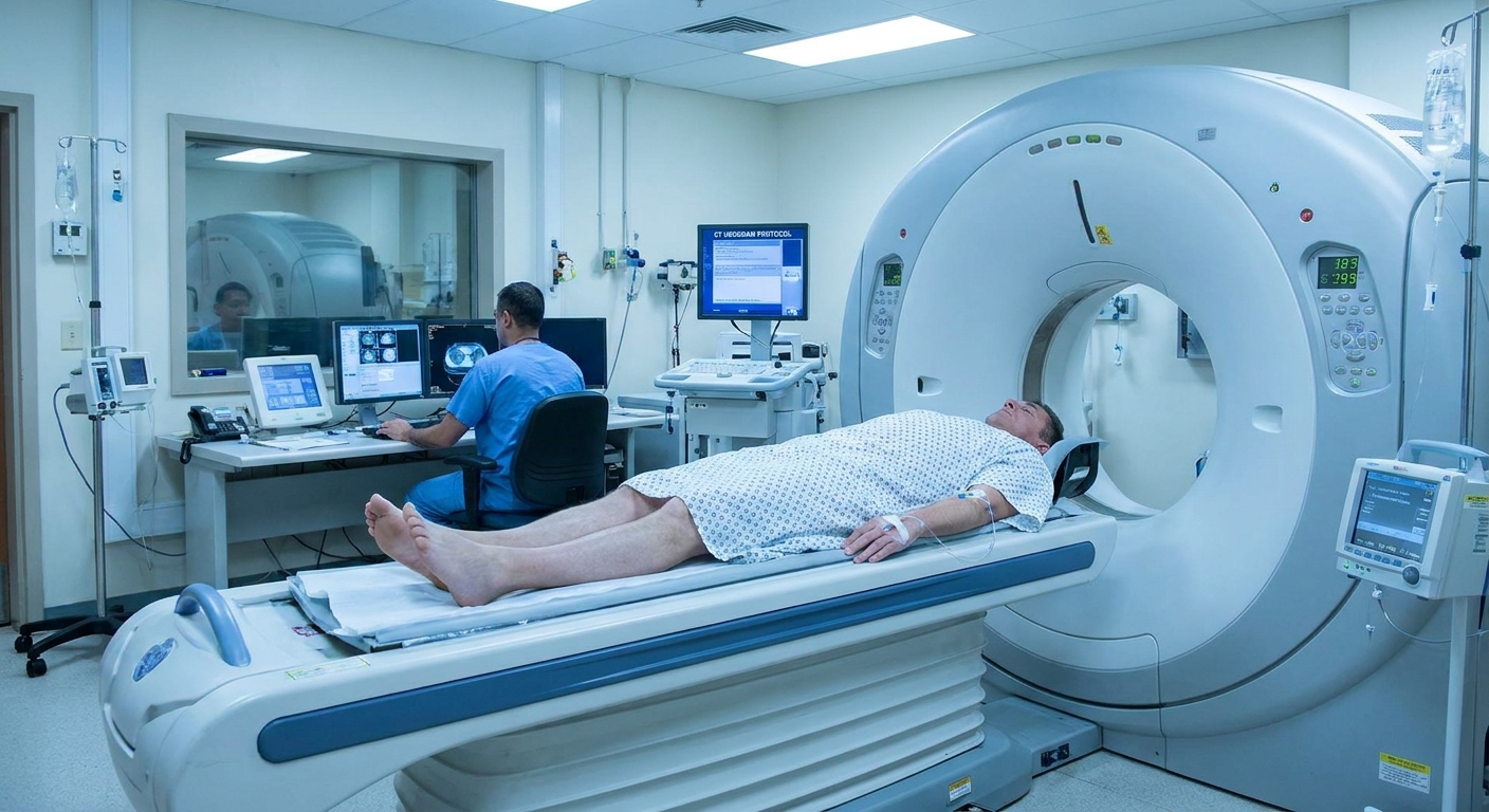
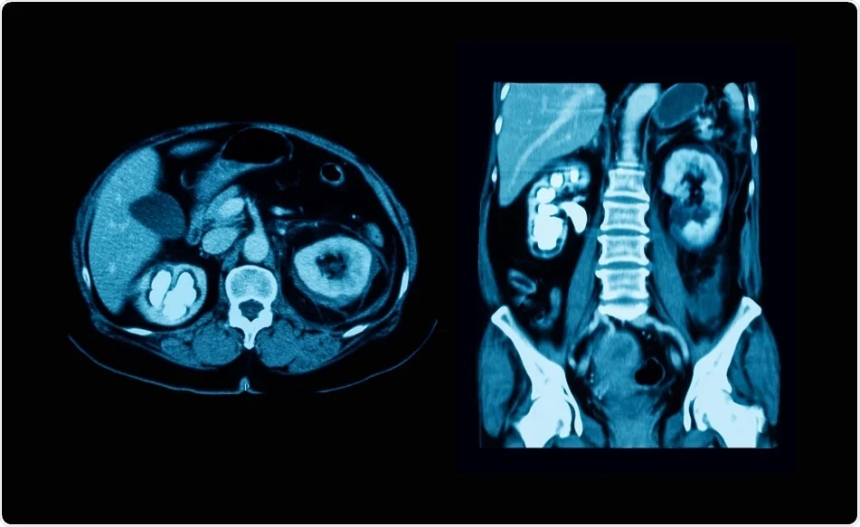
When you live far from specialty care, words on an imaging report can feel like a verdict. I grew up in a rural community, and I still remember how long it could take to get a clear explanation from someone you trust. If your ultrasound or CT report says hydroureter, take a breath. This is a descriptive finding, not a diagnosis by itself. It means the tube that drains urine from the kidney to the bladder, the ureter, looks wider than expected on imaging.
What matters next is why it is dilated and whether there are signs of infection, kidney strain, or a problem at the bladder level.
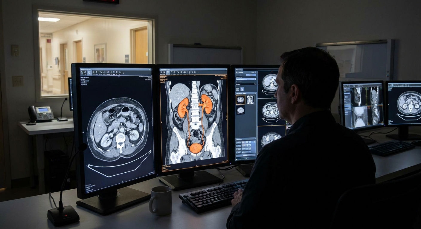
What hydroureter means in plain language
Hydroureter means the ureter is dilated because urine is not draining normally. On a report you might see wording like:
- "Mild right hydroureter"
- "Hydroureter extending to the UVJ"
- "Hydroureter without visible stone"
- "Hydroureteronephrosis" (meaning the ureter is dilated and there is also dilation upstream in the kidney’s drainage system)
A ureter can look dilated for a few reasons:
- Something is blocking urine flow downstream, so urine backs up and stretches the ureter.
- Urine sometimes moves the wrong direction, such as reflux from the bladder toward the ureter.
- The ureter stays widened for a while after a blockage clears, such as after a stone passes.
Hydroureter vs kidney swelling
Hydroureter can appear:
- With kidney swelling upstream, often suggesting urine has had difficulty draining for at least some amount of time.
- Without obvious kidney swelling, which can happen if the issue is mild, intermittent, early, or located low enough that the kidney drainage system has not visibly changed.
If the report mentions hydronephrosis, that refers to dilation in the kidney’s drainage system. You may also see UPJ (ureteropelvic junction), which is where the kidney meets the ureter, and UVJ (ureterovesical junction), which is where the ureter meets the bladder.
Your clinician will interpret these terms in context with your symptoms, lab work, and the rest of the report.
Unilateral vs bilateral hydroureter
One detail that changes the "most likely" list is whether the finding is on one side or both sides:
- Unilateral hydroureter (right or left) often points toward something affecting that one ureter, such as a stone, a stricture, a UVJ issue, or something pressing on that ureter from the outside.
- Bilateral hydroureter more often raises concern for a bladder emptying problem, urinary retention, or bladder outlet obstruction (because both ureters drain into the same bladder).
Common reasons a ureter looks wide on CT or ultrasound
Radiology reports typically point your clinician toward a cause by describing where the ureter is dilated and what is happening near the UVJ and bladder. It also helps to remember that ultrasound may not visualize the entire ureter well, while CT is often better at spotting stones.
1) A stone in the ureter
A very common reason for hydroureter is a ureteral stone, especially when it is lodged in the lower portion of the ureter close to where it enters the bladder. A CT scan often identifies stones well, but very small stones can be missed, and sometimes a report will mention secondary signs such as ureteral dilation or surrounding tissue changes.
Symptoms that often travel with a ureter stone include:
- Sudden, crampy flank or lower abdominal pain that can come in waves
- Pain that may radiate to the groin
- Nausea or vomiting
- Blood in the urine (visible or only on urinalysis)
- Urinary urgency or frequency if the stone is very low
2) Narrowing (stricture) or a structural ureter issue
A ureter can dilate above a narrowed segment called a ureteral stricture. Strictures can develop after prior stones, surgery or instrumentation, inflammation, or radiation. Some people also have structural issues near the UVJ, such as a ureterocele (a ballooning of the ureter as it enters the bladder) that can affect drainage.
3) Blockage at the bladder level or trouble emptying
If the ureter is dilated because urine cannot empty properly into or out of the bladder, the report may point toward a bladder outlet issue or a blockage near where the ureter connects to the bladder.
Potential contributors include:
- Bladder outlet obstruction (for example, from an enlarged prostate, urethral stricture, or significant pelvic organ prolapse)
- Neurogenic bladder or urinary retention, where the bladder does not empty well and pressure backs up
- Mass effect pressing on the bladder neck or UVJ region
Bladder inflammation or infection (cystitis) more often causes burning, urgency, and frequency than it causes a true mechanical blockage. When hydroureter is present, clinicians usually focus on retention and structural obstruction first, while still checking carefully for infection because infection plus obstruction is time-sensitive.
4) External compression in the pelvis or abdomen
Sometimes the ureter is not blocked from the inside, but it is squeezed from the outside. Examples include large fibroids, ovarian cysts or masses, constipation with significant stool burden in some settings, enlarged lymph nodes, and other pelvic or abdominal masses. Some conditions, such as retroperitoneal fibrosis, can also wrap around and narrow a ureter.
5) Reflux: urine washing backward from the bladder
Vesicoureteral reflux means urine can flow backward from the bladder into the ureter, and sometimes higher. This is more common in pediatrics, but it can be seen in adults too, especially with recurrent urinary infections or bladder emptying problems.
Clues that can raise reflux on the list include:
- History of recurrent UTIs, especially febrile kidney infections
- Hydroureter that is intermittent or varies between studies
- Hydroureter without a clear obstructing stone or mass on CT
Reflux is usually confirmed with a specialized test, most often a voiding cystourethrogram (VCUG) or a radionuclide cystogram, rather than a routine ultrasound. In adults, the workup sometimes also includes cystoscopy or bladder function testing depending on the situation.
6) Pregnancy-related dilation
Pregnancy can cause a physiologic hydroureter and hydronephrosis due to hormones and the uterus pressing on the ureters, often more on the right. Because stones and infection can still occur during pregnancy, clinicians interpret this finding carefully alongside symptoms, urine testing, and pregnancy-safe imaging choices.
7) Less common but important causes
Depending on the exact appearance and clinical context, a clinician may also consider ureter or bladder tumors (including urothelial cancer), scarring from prior inflammation, or other rare conditions. This is one reason persistent or unexplained hydroureter often leads to urology follow-up.
How radiologists describe hydroureter
The exact wording on the report can help you ask smarter follow-up questions.
- "Mild" vs "moderate" vs "severe": This is a visual grading, and different radiologists may grade a bit differently. Trend over time matters.
- "To the UVJ" (ureterovesical junction): Suggests the dilation extends down to where the ureter meets the bladder, which can fit with a stone near the bottom, narrowing at the junction, or a bladder-level problem.
- "No obstructing stone seen": The scan did not show a stone blocking flow, but it does not guarantee there is no functional obstruction or that a small stone already passed.
- "Abrupt cutoff": Can suggest a focal blockage, prompting more evaluation depending on severity and symptoms.

What happens next
Hydroureter is often worked up with a combination of symptom review, urine and blood tests, and sometimes repeat imaging.
Tests your clinician may consider
- Urinalysis and urine culture to look for infection and blood
- Kidney function bloodwork, commonly creatinine and estimated GFR
- Repeat ultrasound in a time window that matches your symptoms and risk factors
- CT findings review, especially whether there is a stone, stranding, or a clear transition point
- Additional imaging in selected cases (for example, CT urogram or MRI urogram) when stricture, tumor, or complex anatomy is a concern
Specialty referral
Depending on the full report and your symptoms, your primary care clinician may refer you to urology. That is especially common when there is concern for obstruction, recurrent infections, persistent pain, a single functioning kidney, pregnancy, or worsening kidney function.
When hydroureter is urgent
Hydroureter can become time-sensitive when it is tied to an obstruction plus infection, or when kidney function is at risk.
Seek urgent care or emergency care now if you have hydroureter on imaging and any of the following:
- Fever, chills, or feeling acutely ill, especially with flank pain
- Severe pain that is not controlled with prescribed medications
- Vomiting with inability to keep fluids down
- Little to no urine output or worsening swelling and shortness of breath
- Known pregnancy with significant pain, fever, or urinary symptoms
- One kidney or known chronic kidney disease with new obstruction concerns
If infection is present behind an obstruction, clinicians treat it aggressively because bacteria trapped above a blockage can escalate quickly.
Questions I would ask my clinician
If you have access to your report, these questions often move the conversation forward fast:
- Is the hydroureter on the right, left, or both sides?
- Is there a suspected cause, like a stone, a stricture, or a bladder emptying problem?
- Did the radiologist mention a transition point or "cutoff"?
- Are my kidney function tests normal?
- Do I have signs of infection on urinalysis or culture?
- What symptoms would mean I should go in same day?
- Do you recommend urology, and how soon?
- Do we need repeat imaging to confirm it has resolved?
FAQ
Can hydroureter be temporary?
Yes. A ureter can stay dilated for a period of time after a stone passes or after a short-lived blockage. The timing and the rest of the report help determine whether follow-up imaging is needed.
Does hydroureter always mean something is blocked?
No. Obstruction is common, but it is not the only reason. Reflux, strictures, external compression, and bladder emptying problems can also widen the ureter.
Is hydroureter the same thing as kidney swelling?
No. Hydroureter refers to the ureter being dilated. Your report may or may not mention swelling upstream. If both are present, the report may use combined wording such as hydroureteronephrosis.
What if my report says "mild hydroureter" and I feel fine?
Mild findings sometimes resolve, but they should still be interpreted alongside your history and labs. If you are symptom-free, your clinician may choose watchful waiting with follow-up, or may investigate further if you have risk factors like recurrent infections, reduced kidney function, or persistent findings on repeat imaging.
The takeaway
Hydroureter means the ureter looks dilated on ultrasound or CT. In adults, common causes include stones and other forms of obstruction, but narrowing (stricture), external compression, bladder emptying problems, and less commonly reflux can also be involved. If you have fever, severe pain, vomiting, or difficulty urinating, treat it as urgent. Otherwise, a calm, focused follow-up with your clinician and sometimes urology is usually the right path.