If you have ever read a kidney imaging report and seen the phrase calyceal diverticulum, it can sound alarming. In my clinic days, this was one of those findings patients would circle in red pen and bring to the next visit, understandably worried it meant a tumor or a “hole” in the kidney.
Almost always, a calyceal diverticulum is a benign anatomical pocket. It can still matter, though, because it sometimes becomes a place where urine sits and forms stones or contributes to infection. And occasionally, additional imaging is needed to make sure the finding is truly a diverticulum and not something else that just looks similar on a quick scan.

What it is
A calyceal diverticulum is a small, urine-filled cavity in the kidney that connects to one calyx (a cup-like part of the kidney’s collecting system) through a narrow channel, often called a neck.
Think of your kidney’s collecting system like a set of small funnels (calyces) that drain into a larger funnel (the renal pelvis) and then into the ureter. A calyceal diverticulum is like a little side pocket branching off one funnel. Because the neck is narrow, urine can move in and out slowly, which sets the stage for sediment to settle.
Congenital or acquired?
Classic teaching is that most calyceal diverticula are congenital (present since birth), even if they are not discovered until adulthood. An acquired cause has also been proposed or occasionally reported, such as after inflammation or scarring that affects drainage. Either way, the key feature is the same: it communicates with the collecting system.
Why stones or milk of calcium collect
When urine sits in a low-flow pocket, minerals can settle out. Two classic things radiologists may mention are:
- Stones: Hard mineral deposits that can form within the diverticulum. They may or may not move into the main collecting system.
- Milk of calcium: A layering of tiny calcium crystals suspended in fluid. On imaging, it can look like a smooth, gravity-dependent layer rather than a single solid stone.
Neither of these automatically means you need a procedure. What matters most is whether you have symptoms, recurrent infection, or obstruction.
Symptoms
Many people have a calyceal diverticulum and never feel it. When symptoms happen, they are often related to poor drainage, stones, or infection.
Possible symptoms
- Flank pain (pain in the back or side, usually on one side)
- Recurrent urinary tract infections
- Blood in the urine (visible or microscopic hematuria)
- Kidney stone symptoms, especially if a stone migrates out of the diverticulum
If you have a fever, chills, vomiting, or severe flank pain, treat it as urgent. Infection involving the kidney can become serious quickly.
Diverticulum vs kidney cyst
This is one of the most important distinctions, and it is where confusion often happens on imaging reports.
Calyceal diverticulum
- Connects to the collecting system through a narrow neck
- Contains urine (and sometimes stones or milk of calcium)
- May fill with contrast on delayed imaging because it communicates with urine flow
Simple kidney cyst (including parapelvic cysts)
- Does not connect to the collecting system
- Contains cyst fluid, not urine moving in and out
- Typically behaves like a separate “bubble” in or near the kidney
In plain language: a cyst is usually a sealed sac. A calyceal diverticulum is a pocket that has a doorway into the kidney’s drainage system.
Diverticulum vs hydronephrosis
Hydronephrosis means there is backing up and swelling of the collecting system, often due to a blockage (such as a ureteral stone, scarring, or external compression).
A calyceal diverticulum is different because it is typically:
- Localized: a side cavity off one calyx
- Not the same as global dilation of the renal pelvis and multiple calyces
That said, imaging interpretation can be tricky, especially on ultrasound. If your report mentions “possible diverticulum versus hydronephrosis,” your clinician may recommend additional imaging to clarify.

How it is diagnosed
Calyceal diverticula are usually found on imaging done for another reason, like evaluating pain, infection, or blood in the urine.
Common imaging tests
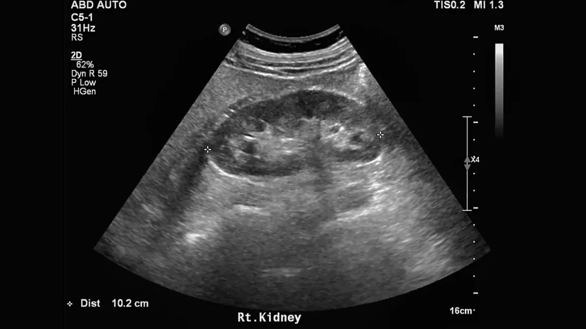
- Ultrasound: May show a cyst-like space, sometimes with echogenic material that layers (suggesting milk of calcium).
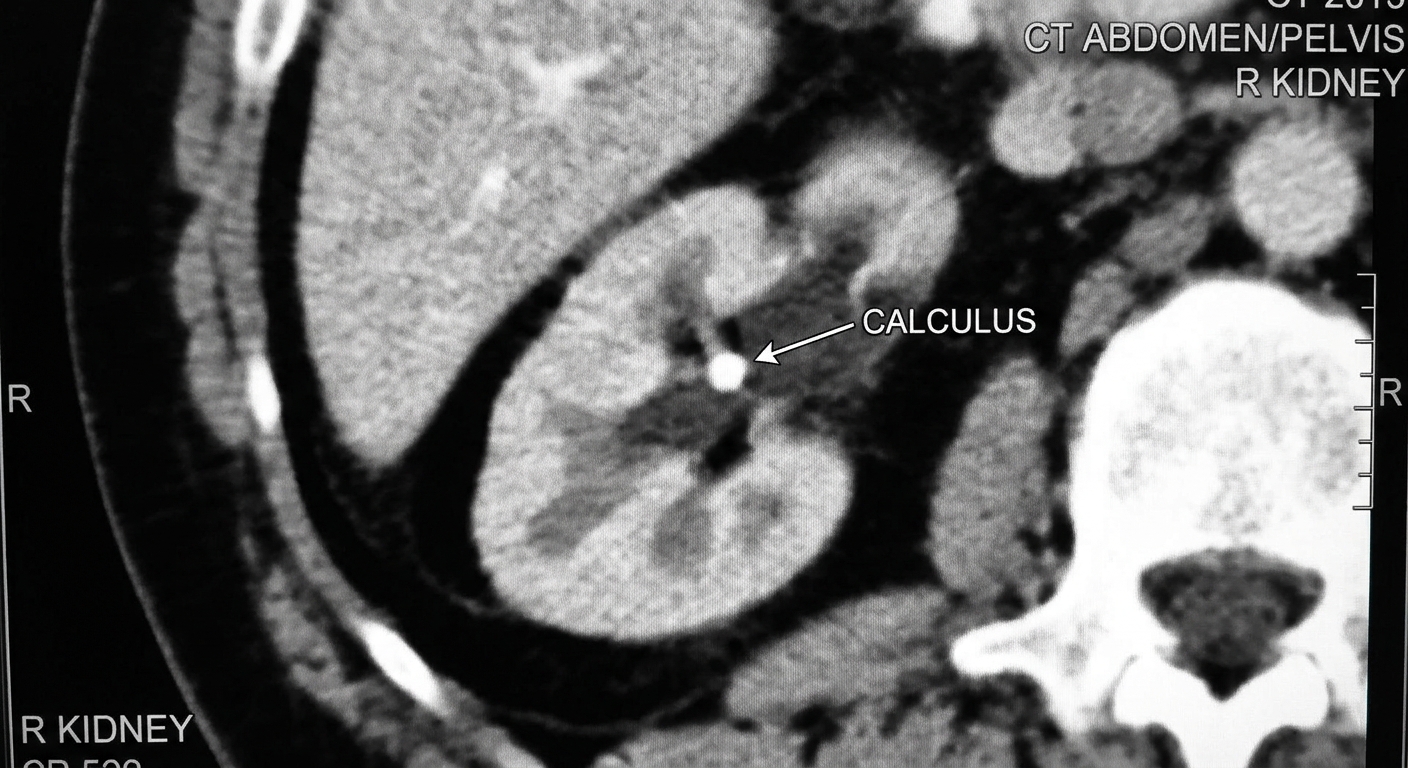
- CT scan: Often shows a cyst-like pocket and can detect stones well. However, a routine CT does not always clearly show the narrow neck.
- CT urography (delayed or excretory-phase CT): This is often the most helpful for confirming a diverticulum because the pocket may fill with excreted contrast if it communicates with the collecting system.
- MRI: Sometimes used when radiation avoidance is important or when CT findings need clarification.
If your report includes terms like “communicates with a calyx,” “narrow neck,” “fills on delayed images,” “milk of calcium,” or “calyceal diverticulum containing calculi,” those are strong clues you are dealing with a diverticulum rather than a simple cyst.
Other look-alikes
Depending on the imaging and the wording of the report, a calyceal diverticulum can be confused with a parapelvic cyst, a hydrocalyx, or focal caliectasis. The deciding feature is usually whether there is communication with the collecting system, especially on delayed contrast imaging.
Treatment
There is no one-size-fits-all approach. Many calyceal diverticula require no treatment and are simply monitored, especially if you have no symptoms.
Often watched when:
- You are symptom-free
- There are no recurrent infections
- Kidney function is stable
- Stones are present but not causing issues
Treatment may be considered when:
- You have recurrent UTIs thought to be related to the diverticulum
- You have persistent or significant flank pain
- There are stones in the diverticulum causing symptoms
- There is concern about poor drainage or repeated inflammation
Urology procedures vary based on the diverticulum’s location (upper, mid, or lower pole), the size of the cavity and neck, and whether stones are present. Common approaches include:
- Ureteroscopy (through the urinary tract) to treat stones and, in selected cases, widen or incise the neck to improve drainage
- Percutaneous approaches (through the skin), which may allow direct stone removal and treatment of the diverticulum cavity in the same setting
- Shockwave lithotripsy in selected cases, though it can be less effective when fragments cannot drain well out of a narrow-neck pocket
Your urologist will match the approach to your anatomy and goals.
Do you need follow-up scans?
Follow-up is often symptom-driven. If you are doing well, your clinician may simply document it and move on. If you have stones, recurrent infections, or ongoing pain, they may recommend repeat imaging to track changes or to better define anatomy, often with a study that includes delayed or excretory images when needed.
Questions to ask
If you are staring at your imaging report and feeling unsure, these questions can help you get a clear plan:
- Does it definitely connect to the collecting system, or could it be a cyst?
- Is there milk of calcium or a true stone inside it?
- Is there any sign of obstruction or hydronephrosis elsewhere?
- Do my symptoms fit this finding, or might something else explain them?
- Do I need follow-up imaging? If so, when and which type?
- Should I see a urologist now or only if symptoms develop?
When to seek urgent care
Contact urgent care or emergency services if you have kidney-related symptoms plus any red flags, including:
- Fever, chills, or feeling acutely ill (possible kidney infection)
- Severe flank pain that does not let up
- Vomiting or inability to keep fluids down
- New confusion or profound weakness, especially with fever or severe dehydration (possible serious systemic illness)
- Blood in the urine with significant pain or clots
These symptoms do not always mean the diverticulum is the cause, but they do mean you should be evaluated quickly.
Bottom line
A calyceal diverticulum is a urine-filled pocket off a single kidney calyx connected by a narrow neck. It is almost always benign, but because urine can stagnate inside, it can become a home for stones or milk of calcium and may contribute to flank pain or recurrent infections.
The most helpful distinction is this: a diverticulum connects to the collecting system, while a simple cyst does not, and hydronephrosis involves backup and dilation of the collecting system rather than a single side pocket. If your report is unclear, it is completely reasonable to ask what feature on imaging confirmed the diagnosis and what, if anything, you need to do next.
FAQ
Is a calyceal diverticulum cancer?
No. A calyceal diverticulum is an anatomic outpouching connected to the collecting system and is not considered a cancer diagnosis. If an imaging report raises concern for a mass, that is a separate conversation and should be clarified directly with your clinician.
Can a calyceal diverticulum turn into hydronephrosis?
Not typically. Hydronephrosis is a dilation of the collecting system from impaired drainage. A diverticulum is a localized side cavity. They are different processes, although both can appear as fluid spaces on imaging.
Can I pass stones from a calyceal diverticulum?
Sometimes. Stones can remain trapped in the diverticulum, or they can move through the narrow neck into the collecting system and then behave like other kidney stones.
Do I need antibiotics?
Antibiotics are used for confirmed or strongly suspected infection, not just because a diverticulum exists. If you have UTI symptoms, fever, or abnormal urine testing, seek medical evaluation.