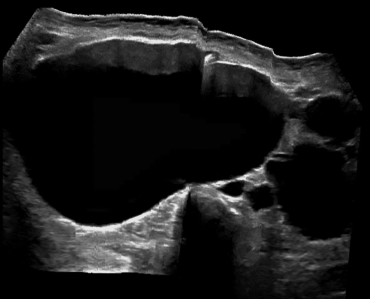
In clinic, I often meet adults who feel blindsided after a scan for kidney stones shows an unfamiliar phrase: medullary sponge kidney. It sounds ominous, but for many people it is a congenital, lifelong kidney structure difference that stays quiet for years and then shows up when stones, urinary tract infections, or blood in the urine appear.
This article walks you through what medullary sponge kidney is, why it is linked to calcium stones, what symptoms to watch for, how it is diagnosed (including the classic “paintbrush” pattern), and how it is different from polycystic kidney disease.

Medullary sponge kidney, simply
Medullary sponge kidney (MSK) is a congenital condition, meaning you are born with it. In MSK, some of the tiny tubes inside the kidney called collecting ducts are wider than usual, especially in the inner part of the kidney known as the medulla.
Think of collecting ducts as small drainage channels that help move urine from the filtering parts of the kidney into the kidney’s drainage system (the calyces and renal pelvis). When these ducts are widened (sometimes described as ectatic), urine can move more slowly through those spaces. Slower flow can encourage:
- Calcium deposits in the inner kidney
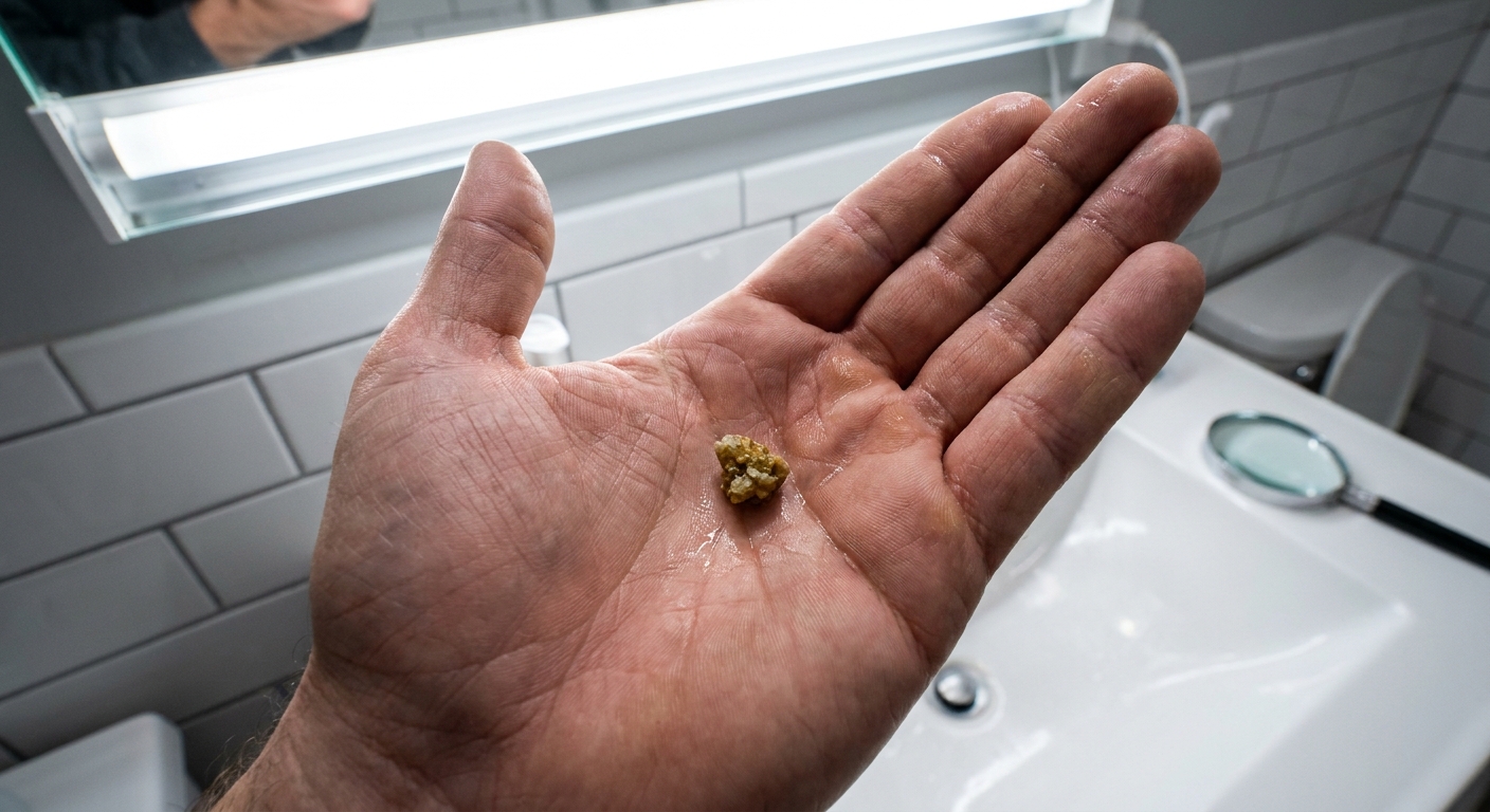
- Kidney stones, especially calcium-based stones
- Recurrent urinary tract infections (UTIs)
- Blood in the urine (hematuria)
Many people with MSK have normal overall kidney function throughout life. A smaller group can develop reduced kidney function over time, most often in the setting of repeated infections, frequent obstruction from stones, or other complicating factors.
Why MSK is linked to calcium stones
The widened collecting ducts can create small pockets where minerals concentrate. Over time, this can lead to calcifications in the medulla, sometimes called medullary nephrocalcinosis.
Common stone types in MSK include calcium oxalate and calcium phosphate. Some people with MSK also have metabolic patterns that add to stone risk, such as:
- Hypercalciuria (too much calcium in the urine)
- Hypocitraturia (too little citrate in the urine, which normally helps prevent stones)
- Distal renal tubular acidosis in some cases, often an incomplete form (a kidney acid-base handling issue that can promote calcium phosphate stones)
If you have MSK and have formed stones, a clinician may recommend a 24-hour urine test to look for these patterns. That information can guide prevention strategies rather than guessing, for example using potassium citrate when citrate is low or urine is too acidic or too alkaline for your stone type.
Symptoms and how MSK is found
Many people do not have symptoms from MSK itself. It is often found incidentally when imaging is done for another reason, or during a workup for stones or recurrent UTIs.
Possible symptoms
- Kidney stone symptoms: flank or back pain, nausea, vomiting, urinary urgency, painful urination
- Blood in the urine: visible pink or red urine, or blood only seen on a urinalysis
- Recurrent UTIs: burning with urination, frequent urination, pelvic discomfort, fever (especially with kidney infection)
When to seek urgent care
Seek prompt evaluation if you have:
- Fever, chills, and flank pain (possible kidney infection)
- Severe pain with vomiting or inability to keep fluids down
- Stone symptoms plus trouble urinating, worsening pain, or you have been told you have a blockage or swelling of a kidney
- Visible blood in the urine, especially with clots
Even if you have MSK, do not assume visible blood in the urine is “just MSK”. Gross hematuria deserves evaluation to rule out infection, a significant stone, and other urinary tract causes.
Diagnosis and the “paintbrush” pattern
MSK is suggested or confirmed primarily by imaging. History and urine or blood tests are most helpful for understanding complications (stones, infections) and for building a prevention plan.
Imaging that may show MSK
- Non-contrast CT: commonly used for suspected stones. It is excellent for detecting stones and medullary nephrocalcinosis, but it does not always show the characteristic collecting-duct pattern because there is no contrast in the urine.
- Excretory-phase contrast imaging: the classic “paintbrush” look is best seen when contrast is excreted into the urine. Historically this was seen on an intravenous pyelogram (IVP). Today, it may also be seen with CT urography or other protocols that include an excretory phase.
- Ultrasound: can show stones, hydronephrosis, and sometimes increased echogenicity from calcifications, but it may miss the finer duct details.
If your report mentions a “paintbrush appearance,” “papillary blush,” or “tubular ectasia,” it is describing that characteristic contrast pattern in the collecting ducts.

MSK vs hydronephrosis
Hydronephrosis means swelling of the kidney’s drainage system (the renal pelvis and calyces) due to urine backing up, often from a blockage like a ureteral stone.
Medullary sponge kidney is different. MSK is a structural widening of inner collecting ducts that you are born with. MSK can contribute to stones, and stones can then cause obstruction and hydronephrosis. But MSK itself is not hydronephrosis.
If your imaging report lists both, it often means: MSK is the background anatomy, and a stone or infection is the current issue.
MSK vs polycystic kidney disease
These two conditions are sometimes confused because both can make kidneys look “abnormal” on imaging, but they are fundamentally different. This is one of those moments where a few precise words in a radiology report really matter.
Medullary sponge kidney
- Congenital widening of collecting ducts in the medulla
- Often associated with calcium stones and medullary calcifications
- Kidney size is often normal
- Kidney function is often preserved
Polycystic kidney disease (PKD)
- Multiple true cysts throughout the kidney tissue (not just widened ducts)
- Kidneys may become enlarged over time
- Can be associated with progressive loss of kidney function
- Often runs strongly in families, especially autosomal dominant PKD
Put plainly: PKD is lots of closed cysts. MSK is widened drainage tubes in the inner kidney, which can look streaky on excretory contrast imaging and tends to show up clinically through stones and infections.
Treatment
There is not a “cure” that changes the kidney anatomy back to typical. Treatment focuses on preventing complications and treating them quickly when they occur. When I talk with patients about MSK, I frame it as a condition you manage by staying ahead of stones and infections, not something you wait to “get worse.”
Preventing kidney stones
- Hydration: Enough fluids to keep urine pale yellow is a simple, powerful step. Many stone formers need higher fluid goals, especially in hot climates or active jobs.
- Diet adjustments: Depending on your stone type and urine testing, your clinician may suggest moderating sodium, keeping dietary calcium appropriate (not zero), and tailoring oxalate intake.
- Medications: Common examples include thiazide-type medications (to reduce urinary calcium) and potassium citrate (to raise urinary citrate and, in selected cases, help correct an incomplete distal renal tubular acidosis pattern). Which medication helps depends on your labs and stone type.
Preventing and treating infections
- Recognize early UTI symptoms and seek testing rather than self-treating repeatedly.
- If UTIs are frequent, ask whether you need an evaluation for stones, urine retention, or other contributors.
- Complete prescribed antibiotics, and ask whether a follow-up culture is needed in your situation.
When stones keep recurring
If you have repeated stones, ask about a full stone-prevention evaluation, including:
- Stone analysis (if you pass or retrieve one)
- Blood work (calcium, kidney function, sometimes parathyroid hormone)
- 24-hour urine testing
It is much easier to prevent the next stone than to manage the pain and disruption of another emergency visit.
Long-term outlook
Most people with medullary sponge kidney do not develop kidney failure from MSK alone. The long-term outlook is usually good, especially when stones and infections are managed proactively.
That said, recurrent kidney infections, repeated obstruction from stones, or extensive calcifications can stress the kidneys over time. Follow-up cadence varies based on your history, but if you have MSK and recurrent stones or UTIs, it is reasonable to ask your clinician what monitoring plan makes sense for you.
Regular check-ins may include:
- Periodic kidney function tests (blood creatinine and estimated GFR)
- Urinalysis when symptoms arise
- Imaging when clinically needed for stones or complications
Common questions
Is medullary sponge kidney genetic?
It is congenital, but in many cases the exact cause is not clear, and it is not inherited in the strong, predictable pattern seen with common forms of polycystic kidney disease. MSK is often sporadic. If multiple family members have stones or unusual kidney findings, it is reasonable to mention it to your clinician.
Can MSK cause pain without a stone?
Most significant kidney pain is tied to obstruction, infection, or inflammation. Some people with MSK report intermittent flank discomfort, but persistent or severe pain deserves evaluation for stones, UTI, or other causes.
Does MSK always show on ultrasound?
Not always. Ultrasound can detect stones, hydronephrosis, and some calcifications, but MSK’s collecting duct changes can be subtle. Non-contrast CT is often better for stones, and excretory-phase contrast imaging is better for showing the classic duct pattern when that detail is needed.
Should I see a nephrologist or a urologist?
It depends on the main issue. Urologists often manage stones that are not passing, obstruction, and procedures. Nephrologists often help with metabolic stone prevention, recurrent nephrocalcinosis, and kidney function monitoring. Many patients benefit from both at different times.
Bottom line
Medullary sponge kidney is a congenital widening of the inner collecting ducts that can set the stage for calcium deposits, kidney stones, UTIs, and blood in the urine. It is not the same condition as polycystic kidney disease, and for most people it does not automatically mean worsening kidney function.
If your scan mentions MSK, the most helpful next step is usually practical: talk with your clinician about stone risk, infection prevention, and whether urine testing could identify a prevention plan tailored to you.